. 2023 Jul 17;6(1):740.
doi: 10.1038/s42003-023-05116-2.

P62/SQSTM1 binds with claudin-2 to target for selective autophagy in stressed intestinal epithelium

Affiliations

P62/SQSTM1 binds with claudin-2 to target for selective autophagy in stressed intestinal epithelium

Rizwan Ahmad et al. Commun Biol. .

Abstract

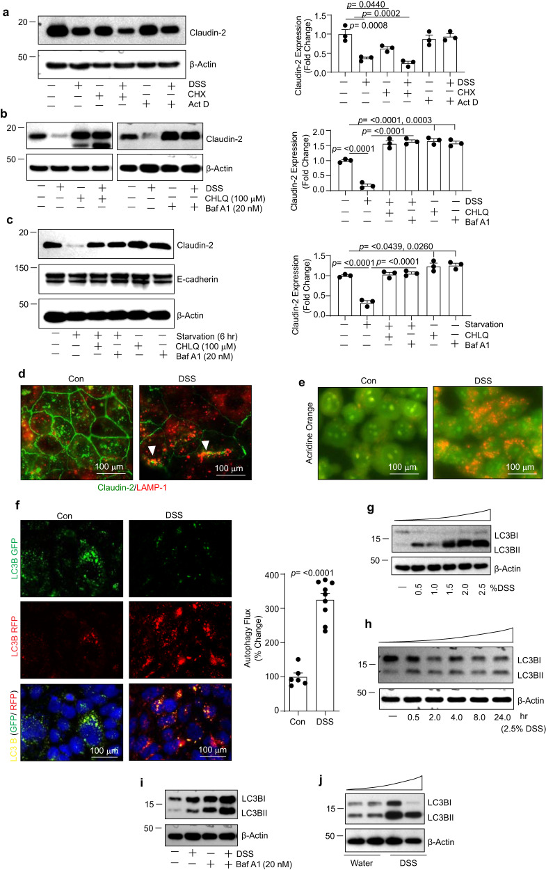
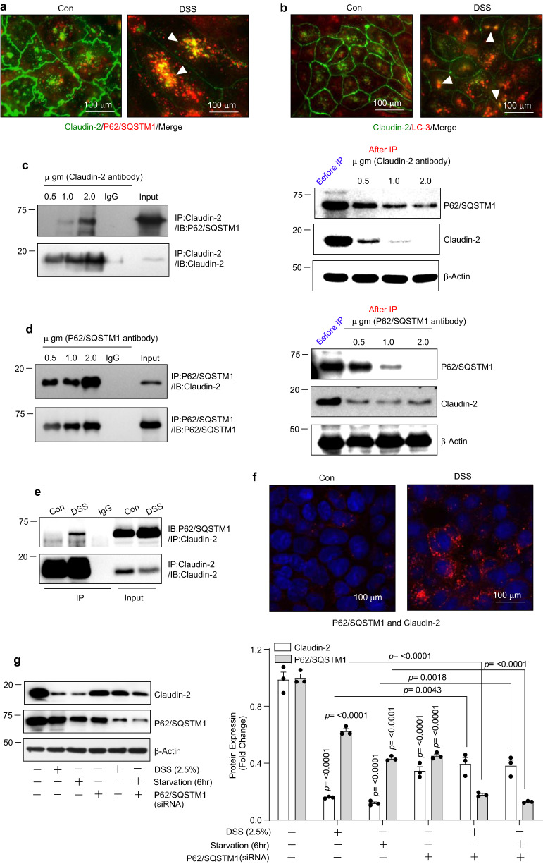
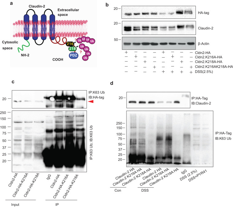
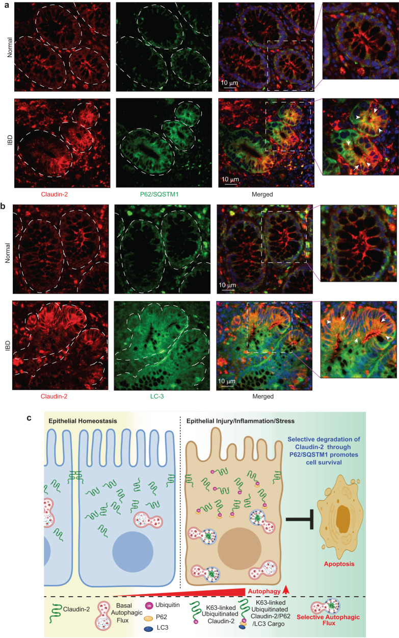
Impaired autophagy promotes Inflammatory Bowel Disease (IBD). Claudin-2 is upregulated in IBD however its role in the pathobiology remains uncertain due to its complex regulation, including by autophagy. Irrespective, claudin-2 expression protects mice from DSS colitis. This study was undertaken to examine if an interplay between autophagy and claudin-2 protects from colitis and associated epithelial injury. Crypt culture and intestinal epithelial cells (IECs) are subjected to stress, including starvation or DSS, the chemical that induces colitis in-vivo. Autophagy flux, cell survival, co-immunoprecipitation, proximity ligation assay, and gene mutational studies are performed. These studies reveal that under colitis/stress conditions, claudin-2 undergoes polyubiquitination and P62/SQSTM1-assisted degradation through autophagy. Inhibiting autophagy-mediated claudin-2 degradation promotes cell death and thus suggest that claudin-2 degradation promotes autophagy flux to promote cell survival. Overall, these data inform for the previously undescribed role for claudin-2 in facilitating IECs survival under stress conditions, which can be harnessed for therapeutic advantages.


Conflict of interest statement

The authors declare no competing interests.

Figures

Fig. 1
Fig. 1. In intestinal epithelial cells, colitogen, or nutrient deprivation (starvation) downregulates claudin-2 expression.
Exposure to DSS inhibited claudin-2 expression in IECs in time- and dose-dependent manners. a Immunoblotting and densitometric analysis using Caco-2 cells and b HT29 cells (n  =  3 independent experiments. Results are presented as means ± SEM. P values from one-way ANOVA followed by Tukey’s post hoc test). c Immunoblotting and densitometric analysis for claudin-2 expression using the lysates from the distal colon from mice subjected to DSS (Acute) colitis (2.5% DSS w/v in drinking water for 7 days; n  =  4 mice/group and results are presented as means ± SEM. P values from Student’s t test). E-cadherin expression served as an epithelial cell marker. d Immunoblotting and densitometric analysis for claudin-2 and claudin-4 expression in Caco-2 cells exposed to TNBS (2.5 mM) for 24-h (n  =  3 independent experiments. Results are presented as means ± SEM. P values from Student’s t test.) in complete culture medium. e Immunoblotting and densitometric analysis for claudin-2 and claudin-4 expression in IECs subjected to nutrient deprivation/starvation (n  =  3 independent experiments. Results are presented as means ± SEM. P values from Student’s t test).
Fig. 2
Fig. 2. Colitogen/stress promote autophagy and claudin-2 protein degradation.
a Immunoblotting and densitometric analysis using Caco-2 cell lysate subjected to DSS treatment with or without co-treatment with Actinomycin-D (Act-D) or Cycloheximide (CHX) (n  =  3 independent experiments. Results are presented as means ± SEM. P values from one-way ANOVA followed by Tukey’s post hoc test). b Immunoblot analysis using total cell lysates. Caco-2 cells were subjected to DSS treatment with or without Chloroquine (100 µM) or Bafilomycin A1 (20 nM). n  =  3 independent experiments. Results are presented as means ± SEM. P values from one-way ANOVA followed by Tukey’s post hoc test. c Immunoblotting and densitometry analysis using total cell lysates of Caco-2 cells subjected to starvation with/without Chloroquine or Bafilomycin A1 (n  =  3 independent experiments. Results are presented as means ± SEM. P values from one-way ANOVA followed by Tukey’s post hoc test). d Representative images of immunofluorescent co-staining for claudin-2 and LAMP1 in control and DSS-treated Caco-2 cells (for 24 h). Arrows indicate colocalization of claudin-2 and LAMP1. e Representative images of Acridine-orange staining in Caco-2 cell treated with DSS (2.5%) (at 24 h post-treatment). f Representative images and autophagy flux rate of LC-3-GFP/RFP reporter construct in control and DSS-treated Caco-2 cells. n  =  3 independent experiments. Results are presented as means ± SEM. P values from Student’s t test. gi Immunoblot analysis for relative expression of LC-3 I and LC-3 II. Lysates from DSS-treated Caco-2 cells (dose- and time-dependent) as well as using Baf A1 pre-treatment were used, respectively. j Immunoblot analysis for relative expression/lipidation of LC-3 I to LC-3 II in colon tissue lysate from mice subjected to acute (DSS) colitis.
Fig. 3
Fig. 3. P62/SQSTM1 physically associates with claudin-2.
a Representative images of immunofluorescent colocalization of claudin-2 and P62/SQSTM1 in control and DSS-treated Caco-2 cells. Arrows indicate colocalization. b Immunofluorescent analysis of claudin-2 and LC-3 expression in control and DSS-treated cells. Arrows indicate colocalization. c Immunoprecipitation of claudin-2 using progressively increasing amount of anti-claudin-2 antibody. The immunoprecipitant and the flow-through samples were immunoblotted for claudin-2 and P62/SQSTM1 proteins. d Immunoblot analysis of claudin-2 and P62/SQSTM1 protein in immunoprecipitant derived using anti- P62/SQSTM1 antibody for immunoprecipitation and using lysates from Caco-2 cell. e Immunoblot analysis for P62/SQSTM1 proteins in immunoprecipitants from control and DSS-treated Caco-2 cell lysates using anti-claudin-2 antibody. f Representative images of proximity-ligation analysis for claudin-2 and P62/SQSTM1 in Caco-2 cells subjected to the DSS treatment. g Immunoblotting and densitometric analysis of claudin-2 expression in Caco-2 cells where P62/SQSTM1 expression was genetically silenced and then cells were subjected to DSS treatment and/or nutrient starvation. n  =  3 independent experiments. Results are presented as means ± SEM. P values from one-way ANOVA followed by Tukey’s post hoc test.
Fig. 4
Fig. 4. Colitogen or nutrient stress induce K63-linked ubiquitination of claudin-2.
a, b Immunoblot analysis of claudin-2 and K63-Ubiquitin using total cell lysate from Caco-2 cells subjected to DSS treatment or starvation with/without PYR41, a cell-permeable ubiquitin E1 ligase inhibitor (5 µM; n  =  3 independent experiments. Results are presented as means ± SEM. P values from one-way ANOVA followed by Tukey’s post hoc test). c Immunoprecipitants pulled down using anti-claudin-2 antibody and Caco-2 cells lysates were immunoblotted using the pan-ubiquitin or claudin-2 antibody. d Immunofluorescent imaging of proximity-ligation assay (PLA) using claudin-2 and Pan-ubiquitin antibody. e Immunoprecipitation was done using Caco-2 cell lysates and antibodies against K63-linked and K48-linked ubiquitin followed by immunoblotting using anti-claudin-2 or P62/SQSTM1 antibody. f Representative images of PLA analysis using antibodies against claudin-2 and K63-Linked ubiquitin in Caco-2 cells subjected to DSS treatment.
Fig. 5
Fig. 5. Lysine (K) 218 in claudin-2 cytoplasmic tail undergoes K63-linked ubiquitination in stressed IECs.
a Schematic representation of lysine positions in the cytoplasmic tail of claudin-2 protein which were substituted with alanine; b Immunoblot analysis using anti-claudin-2 and HA-tag antibodies, and lysates from Caco-2 cell transiently overexpressing Claudin-2-HA(Cldn2-HA), Claudin-2K216A (Cldn2K216A-HA), Claudin-2K218A-HA (cldn2K218A-HA) or both mutations Claudin-2K216AK218A-HA (Cldn2K216AK218A-HA) constructs. Effect of DSS treatment was determined compared to the untreated control cells. c Immunoprecipitation using anti-K63-linked ubiquitin using lysates from Caco-2 cells overexpressing full-length Cldn2-HA or claudin-2 mutant (Cldn2K216A or Cldn2K218A-HA) cDNA constructs. Immunoblotting was done with anti-K63-linked ubiquitin and anti-HA-tag antibody. d Immunoprecipitation using anti-HA-Tag antibody and lysates from Caco-2 cell overexpressing Cldn2-HA, Clan2K216A-HA or Cldn2K218A-HA constructs and treated with DSS. Immunoblotting was done using K63-linked ubiquitin antibodies, respectively.
Fig. 6
Fig. 6. Inhibiting autophagy inhibits claudin-2 protein degradation and promotes cell death in stressed IECs.
a Cell viability in Caco-2 cells subjected to DSS treatment with/without chloroquine and Bafilomycin. n  =  4 independent experiments. Results are presented as means ± SEM. P values from one-way ANOVA followed by Tukey’s post hoc test. b Immunoblotting and densitometric analysis using total colon lysate from WT mice, untreated or treated with 36-077, a potent and selective inhibitor of autophagy. n  =  3 mice/group and results are presented as means ± SEM. P values from Student’s t test. c, d Immunoblotting for ATG16L, claudin-2, and cleaved caspase-3 using lysates from Caco-2 cells transiently transfected with anti-human ATG16L or control siRNA and treated with DSS (2.5%) and starvation. e Cell viability assay using transient transfected claudin-2 siRNA in Caco-2 or HT29 cells stably overexpressing anti-human claudin-2 shRNA, and control cells. n  =  6 independent experiments. Results are presented as means ± SEM. P values from one-way ANOVA followed by Tukey’s post hoc test. f, g Immunoblot analysis for cleaved caspase-3 and claudin-2, and densitometric quantitation. n  =  3 independent experiments. Results are presented as means ± SEM. P values from one-way ANOVA followed by Tukey’s post hoc test. h, i Immunoblot analysis using 3D-cultured freshly isolated mouse colon crypts from WT, Villin-claudin-2 transgenic and claudin-2 knockout mice subjected to DSS treatment (1% DSS w/v), n  =  3 independent experiments. Results are presented as means ± SEM. P values from one-way ANOVA followed by Tukey’s post hoc test. j Representative immunoblots analysis of cleaved caspase-3 expression and densitometry using lysates from Caco-2 cells transiently transfected with full-length or claudin-2 mutant constructs and subjected to DSS treatment. n  =  3 independent experiments. Results are presented as means ± SEM. P values from one-way ANOVA followed by Tukey’s post hoc test.
Fig. 7
Fig. 7. Claudin-2 expression in upregulated in IBD patient samples and co-localizes with autophagy markers.
a Immunofluorescence image of claudin-2 and P62/SQSMT1 using biopsies samples from IBD patients and insert represent the colocalization of claudin-2 and P62/SQSMT1. b Co-immunofluorescence analysis using of anti-claudin-2 and- LC-3 antibodies and insert represent the colocalization of claudin-2 and LC-3. c Graphical representation of the overall concept.

Similar articles

Cited by

References

    1. McGuckin MA, Eri R, Simms LA, Florin TH, Radford-Smith G. Intestinal barrier dysfunction in inflammatory bowel diseases. Inflamm. Bowel Dis. 2009;15:100–113. doi: 10.1002/ibd.20539. - DOI - PubMed
    1. Ahmad R, Sorrell MF, Batra SK, Dhawan P, Singh AB. Gut permeability and mucosal inflammation: bad, good or context dependent. Mucosal Immunol. 2017;10:307–317. doi: 10.1038/mi.2016.128. - DOI - PMC - PubMed
    1. Luster AD, Alon R, von Andrian UH. Immune cell migration in inflammation: present and future therapeutic targets. Nat. Immunol. 2005;6:1182–1190. doi: 10.1038/ni1275. - DOI - PubMed
    1. Zundler S, Becker E, Schulze LL, Neurath MF. Immune cell trafficking and retention in inflammatory bowel disease: mechanistic insights and therapeutic advances. Gut. 2019;68:1688–1700. doi: 10.1136/gutjnl-2018-317977. - DOI - PubMed
    1. Allaire JM, et al. The intestinal epithelium: central coordinator of mucosal immunity. Trends Immunol. 2018;39:677–696. doi: 10.1016/j.it.2018.04.002. - DOI - PubMed

Publication types

LinkOut - more resources