

Cerebral perfusion changes in acute subdural hematoma
- PMID: 37460666
- PMCID: PMC10477107
- DOI: 10.1007/s00701-023-05703-6
Cerebral perfusion changes in acute subdural hematoma
Abstract
Introduction: Acute subdural hematoma (aSDH) is one of the main causes of high mortality and morbidity in traumatic brain injury. Prognosis is poor due to the rapid volume shift and mass effect. Cerebral perfusion is likely affected in this condition. This study quantifies perfusion changes in aSDH using early ER polytrauma CT with perfusion imaging (CTP).
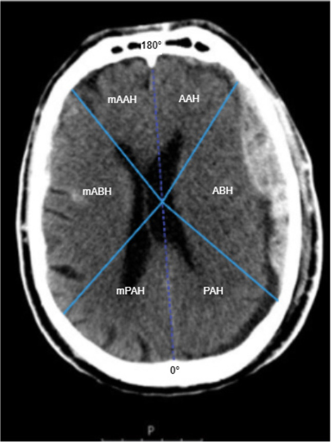
Methods: Data of 54 patients with traumatic aSDH were retrospectively collected. Glasgow Coma scale (GCS), perfusion parameters, therapeutic decisions and imaging data including hematoma thickness, midline shift, and hematoma localization were analyzed. The cortical perfusion parameters of each hemisphere, the area anterior to the hematoma (AAH), area below the hematoma (ABH), area posterior to the hematoma (PAH), and corresponding mirrored contralateral regions were determined.
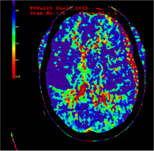
Results: We found a significant difference in Tmax in affected and unaffected whole-hemisphere data (mean 4.0 s vs. 3.3 s, p < 0.05) and a significantly different mean for Tmax in ABH and for the corresponding mirrored area (mABH) (mean 3.8 s vs. 3.1 s, p < 0.05). No significant perfusion changes in cerebral blood flow (CBF), cerebral blood volume (CBV), and mean transit time (MTT) were found.
Conclusion: There was a significant elevation of time to maximum (Tmax) values in the underlying cortical area of aSDH. Possible pathophysiological explanations, the influence on immediate surgical decision-making and further therapeutic consequences have to be evaluated.
Keywords: Acute subdural hematoma; CT perfusion; Cerebral perfusion; Traumatic brain injury.
© 2023. The Author(s).
Conflict of interest statement
The authors declare no competing interests.
Figures

References
-
- Bastos-Leite AJ, Kuijer JP, Rombouts SA, Sanz-Arigita E, van Straaten EC, Gouw AA, van der Flier WM, Scheltens P, Barkhof F. Cerebral blood flow by using pulsed arterial spin-labeling in elderly subjects with white matter hyperintensities. AJNR Am J Neuroradiol. 2008;29:1296–1301. doi: 10.3174/ajnr.A1091. - DOI - PMC - PubMed
Publication types
MeSH terms
LinkOut - more resources
Full Text Sources
