Review
. 2023 Aug;30(8):1869-1885.
doi: 10.1038/s41418-023-01187-0. Epub 2023 Jul 17.

Identity, structure, and function of the mitochondrial permeability transition pore: controversies, consensus, recent advances, and future directions

Affiliations
Review

Identity, structure, and function of the mitochondrial permeability transition pore: controversies, consensus, recent advances, and future directions

Paolo Bernardi et al. Cell Death Differ. 2023 Aug.

Abstract

The mitochondrial permeability transition (mPT) describes a Ca2+-dependent and cyclophilin D (CypD)-facilitated increase of inner mitochondrial membrane permeability that allows diffusion of molecules up to 1.5 kDa in size. It is mediated by a non-selective channel, the mitochondrial permeability transition pore (mPTP). Sustained mPTP opening causes mitochondrial swelling, which ruptures the outer mitochondrial membrane leading to subsequent apoptotic and necrotic cell death, and is implicated in a range of pathologies. However, transient mPTP opening at various sub-conductance states may contribute several physiological roles such as alterations in mitochondrial bioenergetics and rapid Ca2+ efflux. Since its discovery decades ago, intensive efforts have been made to identify the exact pore-forming structure of the mPT. Both the adenine nucleotide translocase (ANT) and, more recently, the mitochondrial F1FO (F)-ATP synthase dimers, monomers or c-subunit ring alone have been implicated. Here we share the insights of several key investigators with different perspectives who have pioneered mPT research. We critically assess proposed models for the molecular identity of the mPTP and the mechanisms underlying its opposing roles in the life and death of cells. We provide in-depth insights into current controversies, seeking to achieve a degree of consensus that will stimulate future innovative research into the nature and role of the mPTP.


Conflict of interest statement

The authors declare no competing interests.

Figures

Fig. 1
Fig. 1. State-of-the-art in mammalian F-ATP synthase purification.
Isolation of mammalian F-ATP synthase from mitochondria is the basis of any in vitro experiment that examines its role as the mPTP. Though difficulties remain, new purification strategies and use of novel detergents have allowed significant progress. Roman numerals I to V indicate selected published examples from refs. [41, 52, 53, 67] and [42], respectively. Micrographs are shown at comparable magnification. SDG sucrose density gradient, SEC size exclusion chromatography.
Fig. 2
Fig. 2. Point mutations of F-ATP synthase affecting its channel properties.
The figure reports mutations of F-ATP synthase that affect specific features of the mPTP, i.e., conductance [47, 58], Ca2+-dependence [98], inhibition by H+ [279], sensitivity to glyoxals [280], and oxidants [281]. For details the reader is referred to the original publications indicated on the picture. Approximate positions of the mutations are indicated by red dots. The structure of F-ATP synthase used in the background is taken from ref. [67].
Fig. 3
Fig. 3. The possible location and conformational changes involved in gating of the mitochondrial F-ATP synthase leak channel.
The diagram was drawn according to recent reports [37, 40, 67, 87, 94]. A Formation of the pore at the F-ATP synthase monomer interface within the dimer. B Reversible opening of the pore within the c-ring under physiological and sub-lethal pathological conditions. Non-reversible dissociation of F1 from FO occurs under severe pathological conditions known to induce cell death. Lipids in the c-ring lumen may be displaced or removed due to the expansion of the c-ring allowing ion conduction. F-ATP synthase subunits are shown as surface representations. Gray arrows indicate the possible path of ion flow through the channel. Brown arrows indicate the putative movements of F1 subcomplex and e-subunit. Figure created with BioRender.com. PDB ID code: 6ZQN was used to illustrate F-ATP synthase and modified for the depiction of the hypothetical conformational changes in channel opening.
Fig. 4
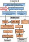
Fig. 4. Possible mechanisms involved in the opening of the mPTP in ischemia reperfusion injury and its prevention by ischemic preconditioning.
This diagram summarizes key factors that have been proposed to initiate and amplify mPTP opening during reperfusion of the heart after a period of prolonged ischemia which itself does not cause mPTP opening because of the low ischemic pHi. Events occurring primarily during ischemia are indicated in pastel blue and those during reperfusion in salmon. The sites at which ischemic preconditioning (IP) may attenuate mPTP opening and hence reduce reperfusion injury (infarct) are indicated with a red minus sign. Further details are given in the text and [123, 182, 190]. This scheme is a modified version of that presented previously [203].

Similar articles

Cited by

References

    1. Haworth RA, Hunter DR. The Ca2+-induced membrane transition in mitochondria. II. Nature of the Ca2+ trigger site. Arch Biochem Biophys. 1979;195:460–7. doi: 10.1016/0003-9861(79)90372-2. - DOI - PubMed
    1. Hunter DR, Haworth RA. The Ca2+-induced membrane transition in mitochondria. III. Transitional Ca2+ release. Arch Biochem Biophys. 1979;195:468–77. doi: 10.1016/0003-9861(79)90373-4. - DOI - PubMed
    1. Hunter DR, Haworth RA. The Ca2+-induced membrane transition in mitochondria. I. The protective mechanisms. Arch Biochem Biophys. 1979;195:453–9. doi: 10.1016/0003-9861(79)90371-0. - DOI - PubMed
    1. Hunter DR, Haworth RA, Southard JH. Relationship between configuration, function, and permeability in calcium-treated mitochondria. J Biol Chem. 1976;251:5069–77. doi: 10.1016/S0021-9258(17)33220-9. - DOI - PubMed
    1. Raaflaub J. Swelling of isolated mitochondria of the liver and their susceptibility to physicochemical influences. Helv Physiol Pharmacol Acta. 1953;11:142–56. - PubMed

Publication types

MeSH terms

Substances