. 2023 Jul 17;13(1):11513.
doi: 10.1038/s41598-023-38696-4.

Glutathione trisulfide prevents lipopolysaccharide-induced retinal inflammation via inhibition of proinflammatory cytokine production in glial cells

Affiliations

Glutathione trisulfide prevents lipopolysaccharide-induced retinal inflammation via inhibition of proinflammatory cytokine production in glial cells

Hiroshi Tawarayama et al. Sci Rep. .

Abstract

We aimed to investigate the impact of glutathione trisulfide (GSSSG) on lipopolysaccharide (LPS)-induced inflammation in retinal glia. Inflammatory responses in mouse-derived glial cells and Wistar rat retinas were stimulated with administration of LPS. Cell survival and proinflammatory cytokine production were examined using the Calcein-AM assay, and reverse transcription-quantitative polymerase chain reaction (RT-qPCR) and enzyme-linked immunosorbent assay (ELISA), respectively. Retinal microglia were visualized with immunohistochemistry for Iba1. Administration of LPS (10 µg/mL) or GSSSG (less than 100 µM) did not affect survival of cultured primary Müller cells and established microglial cells (BV-2). RT-qPCR and ELISA indicated that GSSSG inhibited LPS-induced gene upregulation and protein secretion of proinflammatory cytokines in these glial cells and rat retinas. GSSSG inhibited LPS-induced activation of TGF-β-activated kinase 1 (TAK1), which is an upstream kinase of NF-κB, in BV-2 cells. Finally, in vivo experiments indicated that intravitreal administration of GSSSG but not its relative glutathione disulfide (GSSG) inhibited LPS (500 ng)-induced accumulation of Iba1-immunopositive microglia in rat retinas. Taken together, GSSSG has the potential to prevent pathogenesis of inflammation-associated ocular diseases by inhibiting proinflammatory cytokine expression in retinal glial cells.


Conflict of interest statement

The authors declare no competing interests.

Figures

Figure 1
Figure 1
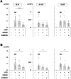
Cell survival of Müller and BV-2 cells treated with GSSSG or LPS. Cell viability of Müller (A) and BV-2 cells (B) treated with various concentrations of GSSSG or LPS for 6 h. Living cells were detected using the Calcein-AM assay, and viability is shown as a percentage of LPS( −)GSSSG( −) controls. **P < 0.01 versus LPS( −)GSSSG( −) (Dunnett's test; n = 4).
Figure 2
Figure 2
Inhibitory effects of GSSSG on LPS-induced expression of proinflammatory cytokines in Müller cells. Changes of IL-6 and Ccl2 mRNA expression (A and B) and protein secretion (C and D) in mouse primary Müller cells. For gene expression analysis using RT-qPCR (A and B), cells were pretreated with GSSSG for 1 h, followed by LPS treatment for 6 h. For protein quantification using ELISA (C and D), cells were also pretreated with GSSSG for 1 h, then subjected to LPS treatment for either 6 h or 24 h. Culture supernatants obtained from cells treated with LPS for 6 h and 24 h were used for protein quantification of IL-6 and Ccl2, respectively. Error bars represent the standard deviation of the mean. ##P < 0.01 versus LPS( −)GSSSG( −) controls (Welch's t test; n = 4); **P < 0.01 versus LPS( +)GSSSG( −) (Dunnett's test; n = 4).
Figure 3
Figure 3
Inhibitory effects of GSSSG on LPS-induced expression of proinflammatory cytokines in BV-2 cells. Changes of TNF-α, Ccl2, IL-6 and IL-1β mRNA expression (A–D) and protein secretion (E–G) in mouse-derived BV-2 cells. For gene expression analysis using RT-qPCR (A-D), cells were pretreated with GSSSG for 1 h, followed by LPS treatment for 6 h. For protein quantification using ELISA (E–G), cells were pretreated with GSSSG for 1 h, followed by LPS treatment for 6 h or 24 h. Culture supernatants obtained from cells treated with LPS for 6 h and 24 h were used for protein quantification of IL-6 (6 h), and TNF-α, Ccl2 and IL-1β (24 h). Error bars represent the standard deviation of the mean. ##P < 0.01 versus LPS( −)GSSSG( −) controls (Welch's t test; n = 4); *P < 0.05 and **P < 0.01 versus LPS( +)GSSSG( −) (Dunnett's test; n = 4).
Figure 4
Figure 4
In vivo effects of GSSSG and GSSG on LPS-induced upregulation of proinflammatory cytokine genes in rat retinas. Changes in IL-6, IL-1β, and Ccl2 mRNA levels in retinas dissected from rat eyes treated with LPS and GSSSG or GSSG. Rats were intravitreally injected with LPS (500 ng) and GSSSG or GSSG (15 or 60 nmol) simultaneously, resulting in putative final glutathione concentrations of 300 µM (A) and 1,200 µM (B) in the vitreous humor. Retinas were collected 10 h after administration. Error bars represent the standard deviation of the mean. ##P < 0.01 versus LPS ( −)GSSSG( −) controls; *P < 0.05, **P < 0.01 (Tukey–Kramer test; n = 12 − 20 in A, 7 − 20 in B. Data for the LPS( −)glutathiones( −) and LPS( +)glutathiones( −) treatment in B are the same as those in A.
Figure 5
Figure 5
Inhibitory effects of GSSSG on LPS-induced accumulation of microglia in rat retinas. (A) Iba1 immunostaining of flat-mounted retinas obtained from LPS- and GSSSG-challenged rats. Rats were intravitreally injected with LPS (500 ng) and GSSSG or GSSG (60 nmol) simultaneously and sacrificed 48 h later. Scale bars: 100 µm. (B) Quantification of Iba1-immunopositive microglia in retinas treated with LPS and GSSSG. Error bars represent the standard deviation of the mean. **P < 0.01 (Tukey–Kramer test; n = 4). n.s.: not significant.
Figure 6
Figure 6
Inhibitory effects of GSSSG on LPS-induced TAK1 phosphorylation in BV-2 cells. (A) Immunoblots of BV-2 cell lysates with antibodies against phosphorylated TAK1 (Ser412) and β-actin as an internal control. BV-2 cells were pretreated with GSSSG (200 µM) for 1 h, followed by treatment with LPS (10 µg/mL) for 30 or 60 min. (B) Quantification of the immunoblot signals. The average was calculated from 4 independent experiments. Error bars represent the standard deviation of the mean. *P < 0.05, **P < 0.01 (Tukey–Kramer test; n = 4).

Similar articles

Cited by

References

    1. Medzhitov R. Inflammation 2010: New adventures of an old flame. Cell. 2010;140:771–776. doi: 10.1016/j.cell.2010.03.006. - DOI - PubMed
    1. Le J, Vilcek J. Interleukin 6: A multifunctional cytokine regulating immune reactions and the acute phase protein response. Lab. Invest. 1989;61:588–602. - PubMed
    1. Hirano T, Akira S, Taga T, Kishimoto T. Biological and clinical aspects of interleukin 6. Immunol. Today. 1990;11:443–449. doi: 10.1016/0167-5699(90)90173-7. - DOI - PubMed
    1. Baggiolini M, Dahinden CA. CC chemokines in allergic inflammation. Immunol. Today. 1994;15:127–133. doi: 10.1016/0167-5699(94)90156-2. - DOI - PubMed
    1. di Giovine FS, Duff GW. Interleukin 1: The first interleukin. Immunol Today. 1990;11:13–20. doi: 10.1016/0167-5699(90)90005-t. - DOI - PubMed

Publication types