. 2023 Oct;18(10):1205-1212.
doi: 10.1038/s41565-023-01443-x. Epub 2023 Jul 17.

DNA-origami-directed virus capsid polymorphism

Affiliations

DNA-origami-directed virus capsid polymorphism

Iris Seitz et al. Nat Nanotechnol. 2023 Oct.

Abstract

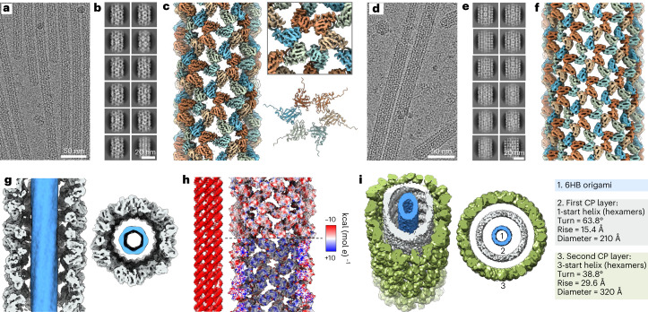
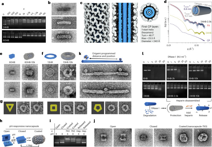
Viral capsids can adopt various geometries, most iconically characterized by icosahedral or helical symmetries. Importantly, precise control over the size and shape of virus capsids would have advantages in the development of new vaccines and delivery systems. However, current tools to direct the assembly process in a programmable manner are exceedingly elusive. Here we introduce a modular approach by demonstrating DNA-origami-directed polymorphism of single-protein subunit capsids. We achieve control over the capsid shape, size and topology by employing user-defined DNA origami nanostructures as binding and assembly platforms, which are efficiently encapsulated within the capsid. Furthermore, the obtained viral capsid coatings can shield the encapsulated DNA origami from degradation. Our approach is, moreover, not limited to a single type of capsomers and can also be applied to RNA-DNA origami structures to pave way for next-generation cargo protection and targeting strategies.


Conflict of interest statement

The authors declare no competing interests.

Figures

Fig. 1
Fig. 1. Formation of capsid-coated DNA origami structures.
a, CPs are isolated from native CCMV (left) and complexed with different DNA origami shapes, resulting in a coating (middle) whose properties are determined by the origami structure. The assembly is driven by electrostatic (positively charged amino acids in the N-terminus marked in red) and protein–protein interactions (right). A second protein layer can develop on top of the first one due to electrostatic interactions between the N-terminus and the negatively charged parts of the CP surface. b, Negative-stain TEM image of plain 6HB structures. c, Negative-stain TEM image of native CCMV particles. d, EMSA shows electrophoretic mobility decrease of 6HB upon complexation with CPs when increasing ε. e, Development of a single layer of CPs on the 6HB origami template using ε ≤ 2k. f, Subsequent development of a second CP layer on top of e. g, Observed size distributions (in diameter) for plain 6HB (blue) and 6HB complexed at ε = 2k (grey) or at ε = 10k (green). The image width of all TEM images corresponds to 500 nm. Source data
Fig. 2
Fig. 2. Single-particle reconstruction of complexed 6HB structures using cryo-EM.
a, Representative micrograph image of 6HB coated with a single protein layer (6HB-2k). b, Selected 2D class averages for 6HB-2k. c, Cryo-EM density of the tube (left) and a selected hexamer (top right). In an atomic model (bottom right) the CP (PDB:1cwp) was flexibly fitted to the EM density. d, Representative image of the double-layered filaments formed by 6HB and the CPs (6HB-10k). e, Selected 2D class averages for 6HB-10k. f, Cryo-EM density of the outer layer of a double-layered tube. g, Cross section and top view of 6HB-2k complexes showing the DNA origami in blue. h, Electrostatic potential surface suggests a negative potential for both the DNA origami (left) and the outward-facing protein surface of the first layer (top right), whereas the DNA adjacent surface of the protein shell (bottom right) possesses a positive potential. i, A 3D model of the assembled double-layer structure shows the different symmetry of the two layers assembling on 6HB origami (1). While the inner layer (2) has a 1-start helix symmetry, the outer layer (3) is defined by a 3-start helix.
Fig. 3
Fig. 3. Single-particle reconstruction of the cap structure.
a, Cryo-EM image of 6HB-2k tubes with caps indicated by white arrows (left) and schematic model of the cap structure (right). b,c, Reconstruction of the cap structure of the first CP layer with fitted models for CP hexamers (H0–H1) and pentamers (P1–P6). b, Side view. c, An oblique view (left) and top view (right) of the cap structure.
Fig. 4
Fig. 4. Applicability of capsid coating on structures with different thickness and shape.
a, EMSA for 24HB and 60HB showing a decrease in the electrophoretic mobility with increasing ε. b, Negative-stain TEM images of plain 24HB and complexed structures with ε = 2.5k and ε = 10k. The image dimensions correspond to 200 nm × 100 nm. c, Cryo-EM density maps for 24HB-2.5k. The origami structure is highlighted in blue. d, SAXS scattering curves measured for CP, 24HB and 24HB-2.5k samples in solution. e, Negative-stain TEM images (125 nm × 125 nm) of plain 60HB and 60HB-10k. f, Negative-stain TEM images (125 nm × 125 nm) of 13HR structure before and after complexation with ε = 10k. g, Complexed 13HR (ε = 10k) can be classified into triangular (left), square-like (middle) and pentagonal (right) shapes. All images have dimensions of 125 nm × 125 nm. h,i, Schematic (h) and EMSA (i) of the different states the nanocapsule can adopt upon pH changes and coating. The coating is initially applied at pH 6 onto the closed nanocapsule, which can be removed by the addition of heparin (Hep). The pH can be changed either once coated or after decoating. j, Negative-stain TEM images of the plain nanocapsule when open, closed and coated (coating applied at pH 6, ε = 750). The image dimensions correspond to 100 nm × 100 nm. k, Application of protein coating on AuNP-functionalized 6HB structures. The image width corresponds to 400 nm. l, Stability of 6HB and 24HB upon DNase I treatment. For both structures, the digestion of the coated DNA origami (single (middle) or double protein layer (right)) is slower than for the plain structures (left). The coating has been removed by heparin before AGE to avoid retention in the wells. Source data
Fig. 5
Fig. 5. Variation of templating material and virus CPs.
a, The RNA–DNA hybrid origami (RNA-6HB) is obtained by thermally annealing 996 nt RNA with DNA staples. The poly(A) tail was left unfolded. b,c, The successful folding can be monitored by AGE (b), in both the ethidium bromide (EtBr) and the Alexa647 (A647) channel, and by AFM (c). d, Folded structures selected from AFM (left) and negative-stain TEM (right) images of RNA-6HB. The image dimensions correspond to 125 nm × 75 nm. e, EMSA for RNA-6HB showing a decrease in the electrophoretic mobility with increasing ε in both the EtBr and the A647 channel. f, Negative-stain TEM images (125 nm × 75 nm) of complexed RNA-6HB structures with ε = 500. g, Observed size distributions (in diameter) for plain RNA-6HB (blue) and RNA-6HB-500 (grey). h, NoVLPs (top, TEM image 150 nm × 150 nm) are disassembled into VP1 units and assembled with 6HB at ε = 500 (bottom). i, VLPs built from the major CP, VP1, of SV40 (top, TEM image 150 nm × 150 nm) are disassembled into their pentameric subunits and reassembled onto 24HB at ε = 5k (bottom, TEM images 200 nm × 100 nm). j, Pentamers formed from the major CP, VP1, of MPyV (top middle, TEM image 150 nm × 150 nm) can either be assembled into VLPs (top left, TEM image 150 nm × 150 nm) or complexed with DNA origami (top right). Development of a pentamer-based protein layer on 6HB (bottom left, TEM images 400 nm × 100 nm) or 24HB (bottom right, TEM images 200 nm × 100 nm) at ε = 1.25k. Source data

Similar articles

Cited by

References

    1. Golub E, et al. Constructing protein polyhedra via orthogonal chemical interactions. Nature. 2020;578:172–176. doi: 10.1038/s41586-019-1928-2. - DOI - PMC - PubMed
    1. Malay AD, et al. An ultra-stable gold-coordinated protein cage displaying reversible assembly. Nature. 2019;559:438–442. doi: 10.1038/s41586-019-1185-4. - DOI - PubMed
    1. Hsia Y, et al. Design of a hyperstable 60-subunit protein icosahedron. Nature. 2016;535:136–139. doi: 10.1038/nature18010. - DOI - PMC - PubMed
    1. Bale JB, et al. Accurate design of megadalton-scale two-component icosahedral protein complexes. Science. 2016;353:389–394. doi: 10.1126/science.aaf8818. - DOI - PMC - PubMed
    1. Marcandalli J, et al. Induction of potent neutralizing antibody response by a designed protein nanoparticle vaccine for respiratory syncytial virus. Cell. 2019;176:1420–1431. doi: 10.1016/j.cell.2019.01.046. - DOI - PMC - PubMed

Publication types