. 2023 Dec;33(12):904-922.
doi: 10.1038/s41422-023-00848-6. Epub 2023 Jul 17.

Mannose antagonizes GSDME-mediated pyroptosis through AMPK activated by metabolite GlcNAc-6P

Affiliations

Mannose antagonizes GSDME-mediated pyroptosis through AMPK activated by metabolite GlcNAc-6P

Yuan-Li Ai et al. Cell Res. 2023 Dec.

Abstract

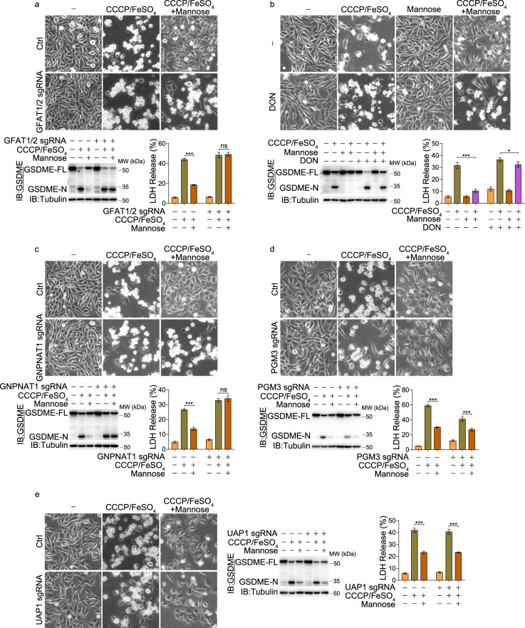
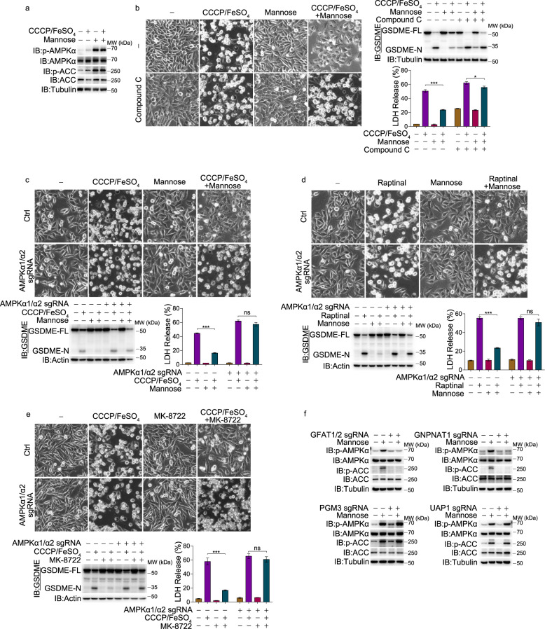
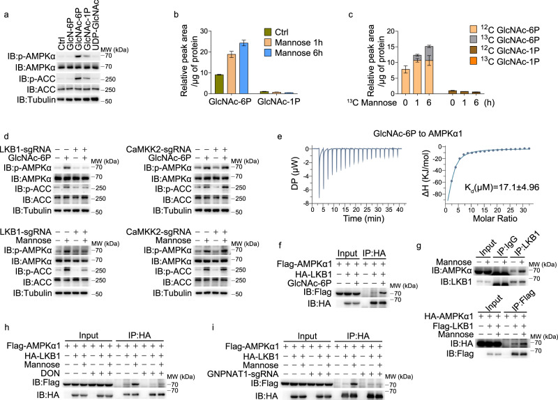
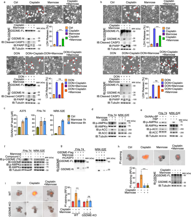
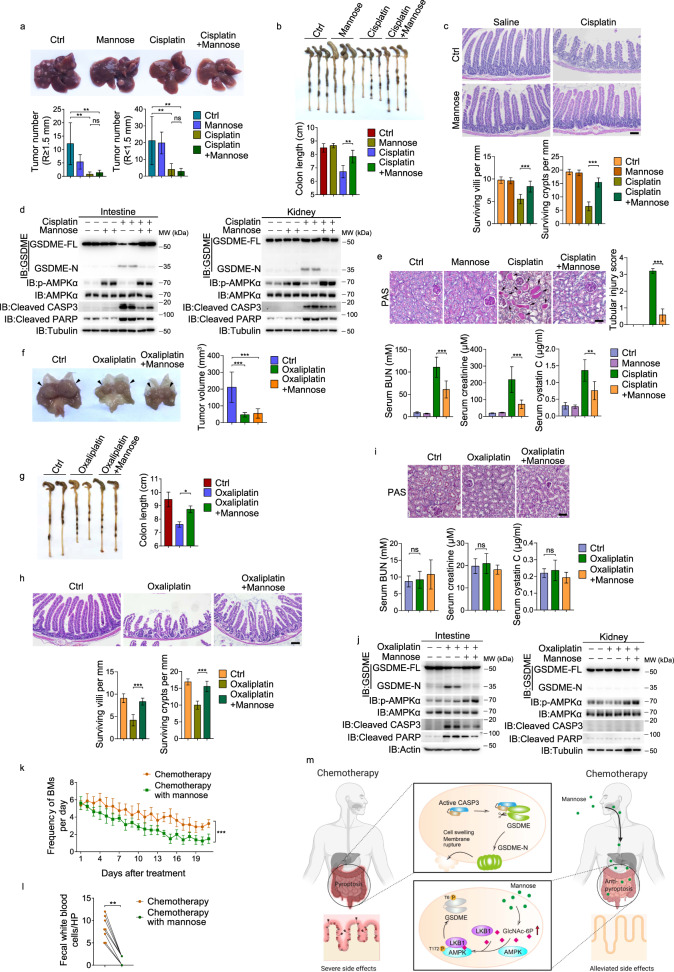
Pyroptosis is a type of regulated cell death executed by gasdermin family members. However, how gasdermin-mediated pyroptosis is negatively regulated remains unclear. Here, we demonstrate that mannose, a hexose, inhibits GSDME-mediated pyroptosis by activating AMP-activated protein kinase (AMPK). Mechanistically, mannose metabolism in the hexosamine biosynthetic pathway increases levels of the metabolite N-acetylglucosamine-6-phosphate (GlcNAc-6P), which binds AMPK to facilitate AMPK phosphorylation by LKB1. Activated AMPK then phosphorylates GSDME at Thr6, which leads to blockade of caspase-3-induced GSDME cleavage, thereby repressing pyroptosis. The regulatory role of AMPK-mediated GSDME phosphorylation was further confirmed in AMPK knockout and GSDMET6E or GSDMET6A knock-in mice. In mouse primary cancer models, mannose administration suppressed pyroptosis in small intestine and kidney to alleviate cisplatin- or oxaliplatin-induced tissue toxicity without impairing antitumor effects. The protective effect of mannose was also verified in a small group of patients with gastrointestinal cancer who received normal chemotherapy. Our study reveals a novel mechanism whereby mannose antagonizes GSDME-mediated pyroptosis through GlcNAc-6P-mediated activation of AMPK, and suggests the utility of mannose supplementation in alleviating chemotherapy-induced side effects in clinic applications.


Conflict of interest statement

The authors declare no competing interests.

Figures

Fig. 1
Fig. 1. Mannose reverses GSDME-mediated pyroptosis in response to different inducers.
Different cancer cell lines were pretreated with mannose (20 mM) for 2 h, followed by different inducers for 24 h to assess pyroptotic features (including characteristic morphology, GSDME cleavage, and LDH release), and the cleaved caspase-3 and its substrate PARP levels were also detected, unless specifically defined. ac Different melanoma cell lines were pretreated with mannose, and then with CCCP/FeSO4 (CCCP, 20 μM; FeSO4, 100 μM). Pyroptosis was detected with morphology (a, red arrows indicate pyroptosis cells), GSDME cleavage (b), and LDH release (c). d Mannose, but not other hexoses, reversed CCCP/FeSO4-induced pyroptotic morphology. Melanoma A375 cells were pretreated with different reagents (20 mM), including glucose, fructose, fucose and galactose for 2 h. e A375 cells were pretreated with mannose, and then with raptinal (1 μM). f, g GSDME-null cancer cell lines PC-9 cells (f) and MDA-MB-468 cells (g) were transfected with GSDME first. The cells were then pretreated with mannose, followed by raptinal. Tubulin or actin was used to determine the amount of loading proteins. All data are presented as mean ± SD of two independent experiments, and one of western blotting results is presented. ***P < 0.001, **P < 0.01.
Fig. 2
Fig. 2. Deletion of genes in HBP impaired the effect of mannose on repressing pyroptosis.
Melanoma A375 cells were pretreated with mannose (20 mM) for 2 h, and then CCCP/FeSO4 (CCCP, 20 μM; FeSO4, 100 μM) for 24 h to assess pyroptotic features (including characteristic morphology, GSDME cleavage, and LDH release), unless specifically defined. a, ce In GFAT1/2 knockout (a), GNPNAT1 knockout (c), PGM3 knockout (d), and UAP1 knockout (e) cells, the effects of mannose on CCCP/FeSO4-induced pyroptosis were evaluated. b Cells were pretreated with mannose and DON (40 μM) for 2 h as indicated, and pyroptosis was determined. Tubulin was used to determine the amount of loading proteins. All data are presented as mean ± SD of two independent experiments, and one of western blotting results is presented. ***P < 0.001, *P < 0.05; ns not significant.
Fig. 3
Fig. 3. Activation of AMPK mediates repression of pyroptosis by mannose.
Melanoma A375 cells were pretreated with mannose (20 mM) for 2 h, and then CCCP/FeSO4 (CCCP, 20 μM; FeSO4, 100 μM) for 6 h to detect AMPK and ACC phosphorylation, or for 24 h to assess pyroptotic features (including characteristic morphology, GSDME cleavage, and LDH release), unless specifically defined. a Cells were treated as described above, and the phosphorylation levels of AMPK (Thr172) and ACC (Ser79) were determined. b Cells were pretreated with compound C (12.5 μM) for 2 h, and pyroptosis was then detected. ce AMPKα1/α2 knockout cells were pretreated with mannose (c, d) or MK-8722 (e, 1 μM) for 2 h, followed by CCCP/FeSO4 (c, e) or raptinal (d), respectively, and pyroptosis was then assayed. f Different genes as indicated were knocked out, respectively, and the phosphorylation levels of AMPK and ACC were determined. Tubulin or actin was used to determine the amount of loading proteins. All data are presented as mean ± SD of two independent experiments, and one of western blotting results is presented. ***P < 0.001, *P < 0.05; ns not significant.
Fig. 4
Fig. 4. GlcNAc-6P directly binds to and activates AMPKα.
a SLO (200 ng/mL) and different metabolites (1 mM) as indicated were incubated with A375 cells for 10 min. The phosphorylation levels of AMPK and ACC were detected. b Intracellular metabolites were extracted from A375 cells, and the peak areas of GlcNAc-6P and GlcNAc-1P were measured by LC-MS. c A375 cells were incubated with 12C6-glucose medium supplemented with 13C6-mannose (20 mM). Intracellular metabolites were extracted and measured by LC-MS. d LKB1 or CaMKK2 knockout A375 cells were incubated with SLO and GlcNAc-6P (top) or treated with mannose (bottom). The phosphorylation levels of AMPK and ACC were detected. e Interaction of purified AMPKα1 and GlcNAc-6P analyzed by ITC. Raw calorimetric data and binding isotherm of the interaction of molecule and protein are shown. f A375 cells were incubated with SLO and GlcNAc-6P for 10 min, and co-IP assay was performed. g A375 cells were treated with mannose for 6 h, and then co-IP assay of endogenous (top) or exogenously transfected (bottom) proteins was performed. h, i In DON-treated (h) or GNPNAT1 knockout (i) A375 cells, co-IP assay was performed. Tubulin was used to determine the amount of loading proteins. All data are presented as mean ± SD of two independent experiments, and one of western blotting results is presented.
Fig. 5
Fig. 5. AMPK phosphorylates GSDME to block caspase-3-mediated cleavage.
Melanoma A375 cells were pretreated with mannose (20 mM) for 2 h, and then CCCP/FeSO4 (CCCP, 20 μM; FeSO4, 100 μM) for 24 h to assess pyroptotic features (including characteristic morphology, GSDME cleavage, and LDH release), and the cleaved caspase-3 and its substrate PARP levels were also detected, unless specifically defined. a Co-IP assay showed the interaction of endogenous AMPK with GSDME. b AMPK directly phosphorylated GSDME in the in vitro AMPK kinase assay. AMPK complex was expressed in HEK293T and purified by HA antibody; GST-GSDME was expressed in E. coli strain BL21 (DE3) and purified by GST pull-down. Reactions were resolved by SDS-PAGE and detected by phospho-antibody. c LC-MS/MS profiling of the in vitro AMPK kinase assay showed that GSDME Thr6 is specifically phosphorylated by AMPK. d Active AMPK was incubated with either GSDME, GSDME T6A or S424A in the in vitro AMPK kinase assay with 32P-labeled ATP. Reactions were resolved by SDS-PAGE and detected by autoradiograph. e Cells were treated with mannose, MK-8722 (1 μM) or metformin (1 mM) for 6 h. GSDME was immunoprecipitated using anti-GSDME antibody, and then the phosphorylation levels of GSDME T6 were detected. f AMPKα1/α2 knockout (left) and compound C-pretreated A375 cells (right) were administered with mannose for 6 h. GSDME was immunoprecipitated using anti-GSDME antibody, and then the phosphorylation levels of GSDME T6 were detected. gh In GSDME knockout cells, GSDME and its point mutant T6E or T6A were separately transfected into cells, and then pyroptosis was detected. i Mannose or MK-8722 (1 μM) were used to treat WT or AMPKα1/α2 knockout A375 cells for 6 h, and the interactions between GSDME and CASP3C/Ap17-HA/p12 were determined. The asterisks indicate co-immunoprecipitated GSDME-Flag. j Cells were transfected with different plasmids as indicated, and the interactions between GSDME mutants and CASP3C/Ap17-HA/p12 were determined. Actin was used to determine the amount of loading proteins. All data are presented as mean ± SD of two independent experiments, and one of western blotting results is presented. ***P < 0.001.
Fig. 6
Fig. 6. Roles of mannose in cisplatin-induced pyroptosis in normal cell lines and organoids.
Normal cell lines were pretreated with mannose (20 mM) for 2 h, and then cisplatin (10 μg/mL) for 24 h to assess pyroptotic features (including characteristic morphology, GSDME cleavage, and LDH release), and the cleaved caspase-3 and its substrate PARP levels were also detected, unless specifically defined. a, b Top, FHs 74 cells (a) and NRK-52E cells (b) were pretreated with mannose and then cisplatin. Bottom, cells were pretreated with mannose and DON (40 μM) and then cisplatin. Pyroptosis was determined. c The level of GlcNAc-6P in cells detected by LC-MS. d Two normal cell lines were treated with mannose with or without DON co-treatment. AMPK and ACC phosphorylation levels were detected. e SLO (200 ng/mL) and metabolite GlcNAc-6P (1 mM) were incubated with cells for 10 min. The phosphorylation levels of AMPK and ACC were detected. FBP was used as a negative control. f Mannose induced GSDME phosphorylation at T6 site. g GSDME was expressed in cells with or without mannose treatment. GSDME proteins were extracted and incubated with reconstituted protein caspase-3. The cleavage of GSDME was detected. The asterisks indicate GSDME-Flag and the arrows indicate cleaved GSDME. h, i Mannose reversed cisplatin-induced pyroptosis (indicated by GSDME cleavage) and pyroptotic cell death (indicated by PI staining) in small intestinal organoids (h) and cisplatin could not induce pyroptotic cell death in GSDME knockout intestinal organoids (i). Tubulin was used to determine the amount of loading proteins. All data are presented as mean ± SD of two independent experiments, and one of western blotting results is presented. ***P < 0.001; ns not significant.
Fig. 7
Fig. 7. Mannose protected organs against chemotherapy drug-induced injury in mouse models.
ac In intestine-specific AMPK-DKO mice (n = 4), mannose lost its effects on protecting small intestine against cisplatin-induced damages, including colon shortening (a), loss of crypts and the reduced villus lengths (b; scale bar, 100 μm), and cleavage of GSDME, caspase-3 and its substrate PARP (c). d Mannose lost its effects on reversing cisplatin-induced pyroptosis (indicated by GSDME cleavage and PI staining) in small intestinal organoids derived from intestine-specific AMPK-DKO mice. eh In GSDMET6E knock-in mice (n = 6), cisplatin lost its effects on inducing damages of small intestine and kidney, including colon shortening (e), loss of crypts and the reduced villus lengths (f; scale bar, 100 μm), damages of kidney (g, top; scale bar, 100 μm), the increased levels of serum BUN, serum creatinine and serum cystatin C (g, bottom), and cleavage of GSDME, caspase-3 and its substrate PARP in both intestine and kidney (h). i Cisplatin was not able to induce pyroptosis (indicated by GSDME cleavage and PI staining) in small intestinal organoids derived from GSDMET6E knock-in mice. Tubulin was used to determine the amount of loading proteins. All data are presented as mean ± SD of two independent experiments, and one of western blotting results is presented. ***P < 0.001, **P < 0.01; ns not significant.
Fig. 8
Fig. 8. Mannose exerts a protective role in small intestine and kidney in mice and patients.
ae DEN/CCl4-inducd HCC mice (n = 5) were treated with cisplatin in the absence or presence of mannose. In liver, the numbers of tumor were counted (a). Mannose rescued cisplatin-induced damages of intestine, including colon shortening (b), loss of crypts and the reduced villus lengths (c; scale bar, 100 μm). Mannose inhibited cisplatin-induced cleavage of GSDME, caspase-3 and its substrate PARP, while elevated AMPK phosphorylation in intestine and kidney (d). Mannose rescued cisplatin-induced damages of kidney, including decreases in the tubular injury score (e, top; scale bar, 100 μm), the levels of serum BUN, serum creatinine, and serum cystatin C (e, bottom). fj Transgenic mice with gastric mucosa-specific expression of COX-2/mPGES-1 (n = 3) were treated with oxaliplatin in the absence or presence of mannose. In hyperplastic stomach, tumor volumes were counted (f). Mannose rescued oxaliplatin-induced damages of intestine, including colon shortening (g), loss of crypts and the reduced villus lengths (h; scale bar, 100 μm). Oxaliplatin induced less damages in kidney (i; scale bar, 100 μm). Mannose inhibited oxaliplatin-induced cleavage of GSDME, caspase-3 and its substrate PARP, while elevated AMPK phosphorylation detected in small intestine and kidney (j). k, l The eight patients received normal chemotherapy with XELOX regimen with or without mannose supplement. Numbers of BMs were recorded daily (k) and fecal WBCs were shown at the end of chemotherapy cycle (l). m A working model for functional mechanism of mannose. Once entering cells, mannose elevates metabolite GlcNAc-6P level, which facilitates interaction of cytosol AMPK and LKB1, leading to AMPK phosphorylation. Activated AMPK phosphorylates GSDME at T6, and results in the failure of GSDME cleavage by caspase-3, thereby inhibiting pyroptotic occurrence. Tubulin or actin was used to determine the amount of loading proteins. All data are presented as mean ± SD of two independent experiments, and one of western blotting results is presented. ***P < 0.001, **P < 0.01, *P < 0.05; ns not significant.

References

    1. Garcia D, Shaw RJ. AMPK: mechanisms of cellular energy sensing and restoration of metabolic balance. Mol. Cell. 2017;66:789–800. doi: 10.1016/j.molcel.2017.05.032. - DOI - PMC - PubMed
    1. Hardie DG, Schaffer BE, Brunet A. AMPK: an energy-sensing pathway with multiple inputs and outputs. Trends Cell Biol. 2016;26:190–201. doi: 10.1016/j.tcb.2015.10.013. - DOI - PMC - PubMed
    1. Olivier S, Foretz M, Viollet B. Promise and challenges for direct small molecule AMPK activators. Biochem. Pharmacol. 2018;153:147–158. doi: 10.1016/j.bcp.2018.01.049. - DOI - PubMed
    1. Treebak JT, Birk JB, Hansen BF, Olsen GS, Wojtaszewski JF. A-769662 activates AMPK beta1-containing complexes but induces glucose uptake through a PI3-kinase-dependent pathway in mouse skeletal muscle. Am. J. Physiol. Cell Physiol. 2009;297:C1041–C1052. doi: 10.1152/ajpcell.00051.2009. - DOI - PubMed
    1. Zhang CS, et al. Fructose-1,6-bisphosphate and aldolase mediate glucose sensing by AMPK. Nature. 2017;548:112–116. doi: 10.1038/nature23275. - DOI - PMC - PubMed

Publication types