. 2023 Aug;5(8):1290-1302.
doi: 10.1038/s42255-023-00835-6. Epub 2023 Jul 17.

Fatty acid oxidation organizes mitochondrial supercomplexes to sustain astrocytic ROS and cognition

Affiliations

Fatty acid oxidation organizes mitochondrial supercomplexes to sustain astrocytic ROS and cognition

Brenda Morant-Ferrando et al. Nat Metab. 2023 Aug.

Abstract

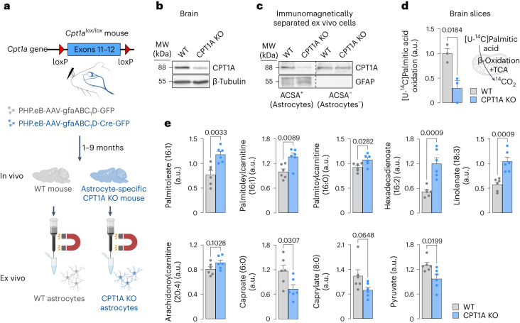
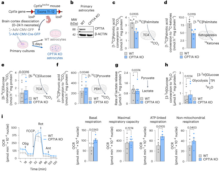
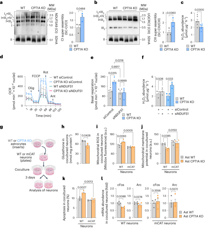
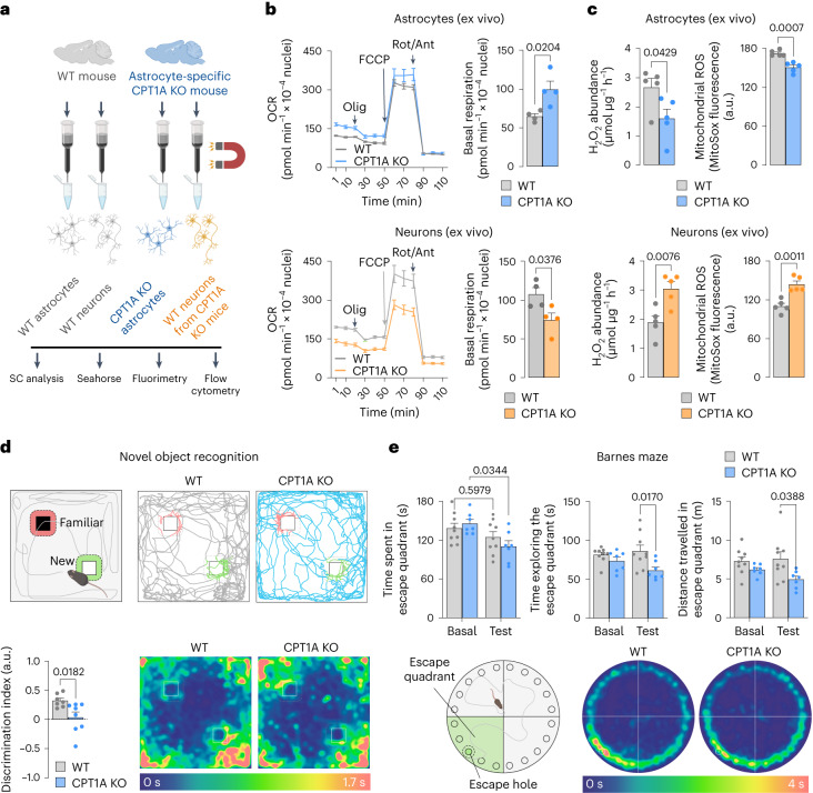
Having direct access to brain vasculature, astrocytes can take up available blood nutrients and metabolize them to fulfil their own energy needs and deliver metabolic intermediates to local synapses1,2. These glial cells should be, therefore, metabolically adaptable to swap different substrates. However, in vitro and in vivo studies consistently show that astrocytes are primarily glycolytic3-7, suggesting glucose is their main metabolic precursor. Notably, transcriptomic data8,9 and in vitro10 studies reveal that mouse astrocytes are capable of mitochondrially oxidizing fatty acids and that they can detoxify excess neuronal-derived fatty acids in disease models11,12. Still, the factual metabolic advantage of fatty acid use by astrocytes and its physiological impact on higher-order cerebral functions remain unknown. Here, we show that knockout of carnitine-palmitoyl transferase-1A (CPT1A)-a key enzyme of mitochondrial fatty acid oxidation-in adult mouse astrocytes causes cognitive impairment. Mechanistically, decreased fatty acid oxidation rewired astrocytic pyruvate metabolism to facilitate electron flux through a super-assembled mitochondrial respiratory chain, resulting in attenuation of reactive oxygen species formation. Thus, astrocytes naturally metabolize fatty acids to preserve the mitochondrial respiratory chain in an energetically inefficient disassembled conformation that secures signalling reactive oxygen species and sustains cognitive performance.


Conflict of interest statement

The authors declare no competing interests.

Figures

Fig. 1
Fig. 1. In vivo astrocyte-specific Cpt1a KO inhibits fatty acid oxidation and alters the metabolomics pattern in the brain.
a, Strategy used to generate astrocyte-specific Cpt1a KO mice and to immunomagnetically purify CPT1A KO astrocytes from adult brain. Created with BioRender.com. b, Western blot against CPT1A protein in astrocyte-specific Cpt1a KO brain. β-Tubulin was used as a loading control; n = 2 mice per condition (Supplementary Fig. 1d). c, Western blotting against CPT1A protein in ACSA+ (astrocytes) and ACSA (not astrocytes) cells, immunomagnetically isolated from astrocyte-specific Cpt1a KO mouse brain; n = 2 mice per condition. GFAP was used as astrocyte enrichment and loading controls. d, Rate of 14CO2 production from [U-14C]palmitic acid in brain slices of WT and astrocyte-specific Cpt1a KO mice. Data are mean ± s.e.m. P value is indicated (n = 3 biologically independent samples; unpaired Student’s t-test, two-sided). e, Concentrations of a selection of metabolites altered in the metabolomics study of the brain samples from astrocyte-specific Cpt1a KO when compared with WT mice. Data are mean ± s.e.m. P values are indicated (n = 6 mice per condition; unpaired Student’s t-test, two-sided). a.u., arbitrary units. Source data
Fig. 2
Fig. 2. KO of Cpt1a in astrocytes inhibits fatty acid oxidation and metabolic rewiring enhancing mitochondrial oxygen consumption.
a, Strategy used to obtain Cpt1a KO astrocytes in astrocytes in primary culture. Created with BioRender.com. b, Western blot against CPT1A protein in Cpt1a KO astrocytes in primary culture 5 days after AdV-CMV-Cre-GFP transduction; n = 3 biologically independent cell culture preparations; unpaired Student’s t-test, two-tailed. β-Actin was used as a loading control (Supplementary Fig. 2b). c, 14CO2 production from [1-14C]palmitic acid in WT and Cpt1a KO astrocytes in primary culture. Data are mean ± s.e.m. P value is indicated (n = 4 biologically independent samples; unpaired Student’s t-test, two-sided). d, 14Ketones production from [1-14C]palmitic acid in WT and Cpt1a KO astrocytes in primary culture. Data are mean ± s.e.m. P value is indicated (n = 4 biologically independent samples; unpaired Student’s t-test, two-sided). eh, 14CO2 production from [6-14C]glucose (e) or [1-14C]pyruvic acid (f), rate of lactate released (g) and glycolytic flux as measured by the rate of [3-3H]glucose conversion into 3H2O (h), in WT and Cpt1a KO astrocytes in primary culture. TPI, triosephosphate isomerase. Data are mean ± s.e.m. P values are indicated; n = 6 (e), 6 (f), 8 (g) and 6 (h) biologically independent cell culture preparations; paired Student’s t-test, two-sided. i, OCR analysis and calculated parameters in WT and Cpt1a KO astrocytes in primary culture. Data are mean ± s.e.m. P values are indicated (n = 5 biologically independent cell culture preparations; unpaired Student’s t-test, two-sided) (Supplementary Fig. 2j,k). Source data
Fig. 3
Fig. 3. KO of Cpt1a in astrocytes induces mitochondrial SCs leading to increased respiration and decreased ROS affecting bioenergetics and function of cocultured neurons.
a, Free CI and CI-containing SCs (SC-CI) in WT and Cpt1a KO primary astrocytes, analysed by BNGE followed by immunoblotting against CI subunit NDUFA9. Data are mean ± s.e.m. P values are indicated (n = 3 biologically independent cell culture preparations; unpaired Student’s t-test, two-sided). b, Free CIII and CIII-containing SCs (SC-CIII) in WT and Cpt1a KO primary astrocytes, analysed by BNGE followed by immunoblotting against CIII subunit UQCRC2. Data are mean ± s.e.m. P values are indicated (n = 3 biologically independent cell culture preparations; unpaired Student’s t-test, two-sided). c, H2O2 production in WT and CPT1A KO astrocytes in primary culture. Data are mean ± s.e.m. P values are indicated (n = 6 independent cell culture preparations; unpaired Student’s t-test, two-sided). d, OCR analysis in WT and Cpt1a KO astrocytes in primary culture, either transfected with scrambled (control) or NDUFS1 siRNAs. Data are mean ± s.e.m. P values are indicated (n = 4 biologically independent cell culture preparations) (Supplementary Fig. 3f). e, Basal respiration in WT and CPT1A KO astrocytes in primary culture, either transfected with scrambled (control) or NDUFS1 siRNAs. Data are mean ± s.e.m. P values are indicated (n = 4 biologically independent cell culture preparations; two-way ANOVA followed by Tukey). f, H2O2 production by WT and CPTA1A KO astrocytes in primary culture, either transfected with scrambled (control) or NDUFS1 siRNAs. Data are mean ± s.e.m. P values are indicated (n = 3 biologically independent cell culture preparations; multiple unpaired Student’s t-test). g, Strategy used to assess the effect of Cpt1a KO astrocytes on WT or mCAT neurons in primary culture. Created with BioRender.com. hl, Glutathione concentration (h), mitochondrial ROS (i), ∆ψm (j), apoptosis (k) and c-Fos and Arc mRNA abundances (l) in WT or mCAT-expressing transgenic neurons after coculture with WT or Cpt1a KO astrocytes; n = 3 (h), 4 (i), 4 (j), 4 (k, WT), 4 (k, mitoCAT) and 4 (l) biologically independent cell culture preparations; paired Student’s t-test, two-sided for simple comparisons and two-way ANOVA followed by Tukey for multiple comparisons. Source data
Fig. 4
Fig. 4. Astrocyte-specific Cpt1a KO mice enhance in astrocytes but decrease in neurons mitochondrial SCs and respiration causing cognitive impairment.
a, Strategy used to immunomagnetically isolate astrocytes and neurons from WT or astrocyte-specific CPT1A KO adult mice. Created with BioRender.com. b, OCR analysis and calculated basal respiration in immunomagnetically isolated astrocytes (top) and neurons (bottom) from WT and astrocyte-specific Cpt1a KO mice. Data are mean ± s.e.m. P values are indicated (n = 4 mice per genotype; unpaired Student’s t-test, two-sided). c, H2O2 and mitochondrial ROS analyses in immunomagnetically isolated astrocytes (top) and neurons (bottom) from WT and astrocyte-specific Cpt1a KO mice. Data are mean ± s.e.m. P values are indicated (n = 5 mice per genotype; unpaired Student’s t-test, two-sided). d, Novel object recognition test in WT and astrocyte-specific Cpt1a KO mice. Representative paths and spatiotemporal quantitative heatmaps are shown. Data are mean ± s.e.m. P values are indicated (n = 9 (WT) or 7 (CPT1A KO) mice; unpaired Student’s t-test, two-sided). e, Barnes maze test in WT and astrocyte-specific Cpt1a KO mice 8 days after training. Spatiotemporal quantitative heatmaps are shown. Data are mean ± s.e.m. P values are indicated (n = 9 (WT) or 7 (CPT1A KO) mice; two-way ANOVA followed by Tukey). P values in the figure. Source data

References

    1. Hosli, L. et al. Direct vascular contact is a hallmark of cerebral astrocytes. Cell Rep.39, 110599 (2022). 10.1016/j.celrep.2022.110599 - DOI - PubMed
    1. Kacem, K., Lacombe, P., Seylaz, J. & Bonvento, G. Structural organization of the perivascular astrocyte endfeet and their relationship with the endothelial glucose transporter: a confocal microscopy study. Glia23, 1–10 (1998). 10.1002/(SICI)1098-1136(199805)23:1<1::AID-GLIA1>3.0.CO;2-B - DOI - PubMed
    1. Almeida, A., Moncada, S. & Bolaños, J. P. Nitric oxide switches on glycolysis through the AMP protein kinase and 6-phosphofructo-2-kinase pathway. Nat. Cell Biol.6, 45–51 (2004). 10.1038/ncb1080 - DOI - PubMed
    1. Barros, L. F., Porras, O. H. & Bittner, C. X. Why glucose transport in the brain matters for PET. Trends Neurosci.28, 117–119 (2005). 10.1016/j.tins.2005.01.002 - DOI - PubMed
    1. Magistretti, P. J. & Allaman, I. A cellular perspective on brain energy metabolism and functional imaging. Neuron86, 883–901 (2015). 10.1016/j.neuron.2015.03.035 - DOI - PubMed

Publication types