. 2023 Jun;59(6):395-400.
doi: 10.1007/s11626-023-00778-y. Epub 2023 Jul 17.

Establishment of induced pluripotent stem cells derived from patients and healthy siblings of a nevoid basal cell carcinoma syndrome family

Affiliations

Establishment of induced pluripotent stem cells derived from patients and healthy siblings of a nevoid basal cell carcinoma syndrome family

Yoji Nakase et al. In Vitro Cell Dev Biol Anim. 2023 Jun.

Abstract

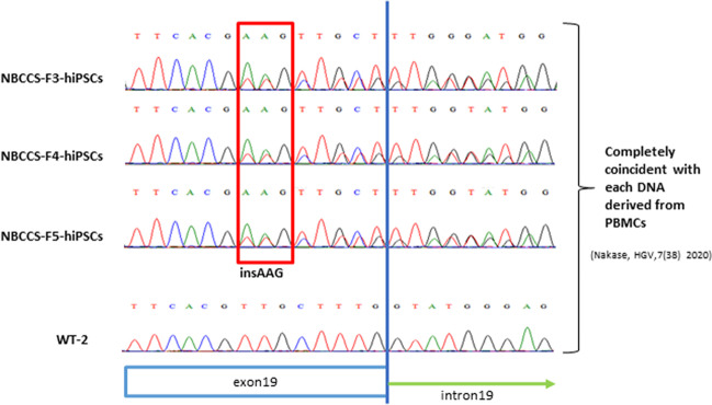
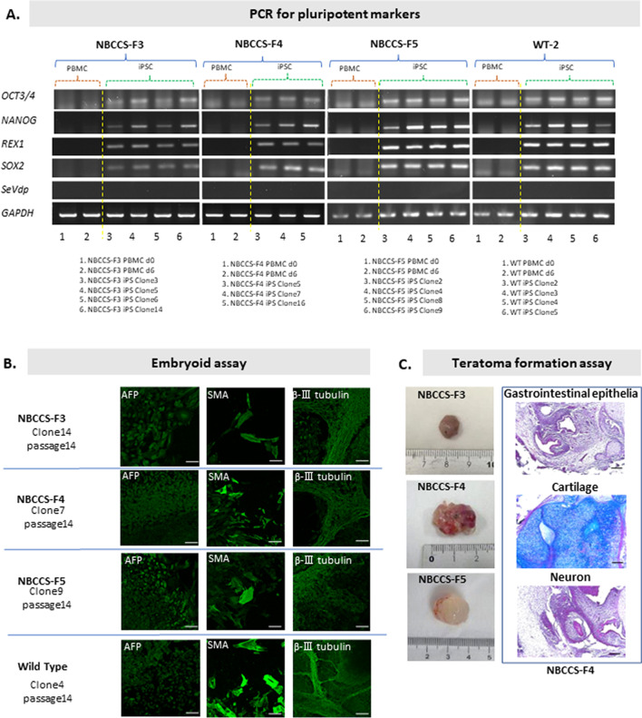
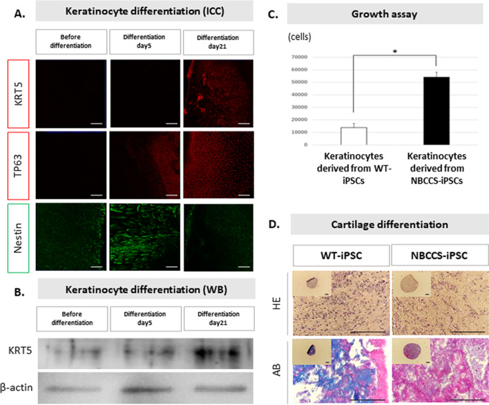
It is known that a nevoid basal cell carcinoma syndrome (NBCCS) is characterized by a combination of developmental abnormalities and a predisposition to form various tumors. Although it is possible to create disease models via gene editing, there are significant potential problems with this approach such as off-target mutations and differences in SNPs. On the other hand, since disease families share common SNPs, research using iPSCs derived from both patients and healthy siblings of the same disease family is very important. Thus, establishment of induced pluripotent stem cells derived from patients and healthy siblings of the same NBCCS family will be of great importance to study the etiology of this disease and to develop therapeutics. In this study, we generated hiPSCs using peripheral blood mononuclear cells derived from the patients and healthy siblings of familial NBCCS with the novel mutation in PTCH1_c.3298_3299insAAG in the feeder- and serum-free culture conditions using SeVdp. In addition, disease-specific hiPSCs such as those expressing the PTCH1_c.3298_3299insAAG mutation could be powerful tools for revealing the genotype-phenotype relationship and pathogenicity of NBCCS.


Figures

Figure 1.
Figure 1.
PTCH1 mutation of familial NBCCS-hiPSCs. The purified polymerase chain reaction (PCR) products of PTCH1 exon19 were screened by Sanger sequencing using the PowerPlex 16 system (Promega Corporation, Madison, WI), and analyses were performed with the ABI PRISM 3100 Genetic analyzer and Gene Mapper v3.5 (Applied Biosystems). The results of direct sequencing of PTCH1 exon 19 are shown. Though an insertion was not detected in WT-hiPSC, an AAG insertion was detected between coding sequences 3298 and 3299 in NBCCS-F4-, F5-, and F6-hiPSC.
Figure 2.
Figure 2.
Assay of pluripotency and differentiation ability of NBCCS-hiPSCs. All procedures are performed under feeder-, serum-, and integration-free conditions. Briefly, after separating and culturing PBMCs in serum-free RD6F (Sato et al. 1987) supplemented with IL-2 (CELEUK; Takeda Pharm., Osaka, Japan) (Okamoto et al. 1996), for 6 d, PBMCs were reprogrammed with SeVdp (KOSM) 302 L (Nishimura et al. 2017), which does not integrate into the host genomic background, and further cultured in hESF9 serum-free medium on Laminin-E8 (Nippi, Tokyo, Japan)-coated dish (Hamada et al. 2020a). (A) PCR for pluripotent makers was performed with TRIzol RNA Isolation Reagents (Thermo Scientific, Waltham, MA), RNA-to-cDNA master mix (Applied Biosystems, Carlsbad, CA), KOD-FX Neo (Toyobo, Osaka, Japan), and ChemiDoc Touch Imaging System (Bio-Rad, Hercules, CA). (B) To confirm the differentiation ability of the hiPSCs, embryoid assay and teratoma formation assay were performed. Briefly, for embryoid assay, undifferentiated NBCCS-hiPSCs were cultured in a low-attachment dish, in which gathered sphere formed embryoids. The embryoids were transferred onto a gelatin-coated dish and cultured for 3 wk. After differentiation, immunocytochemistry (ICC) was performed, and fluorescence images were captured using a Zeiss inverted LSM 700 confocal microscope (Carl Zeiss GmbH, Jena, Germany). (C) For teratoma formation assay, undifferentiated NBCCS-hiPSCs were injected into the dorsal flank of SCID (CB17/Icr-Prkdcscid/CrlCrlj) mice, and dissected tumors were stained with hematoxylin/eosin (HE) and AB, and histologically analyzed using a Nikon ECLIPSE E800 microscope (Nikon Corporation, Tokyo, Japan) and photographed with a Leica DC500 camera (Leica Microsystems AG, Wetzlar, Germany). Each bar indicates 100-µm length.
Figure 3.
Figure 3.
Disease modeling of NBCCS using NBCCS-hiPSCs. (A ~ C) For keratinocyte differentiation (Kogut I, 2014), NBCCS-iPSCs were seeded at 1 × 105 cells/well in a 35-mm dish coated with Laminin-E8, cultured in hESF9 medium with Rock inhibitor (10 μM) (Y-27632, Wako, Osaka, Japan) for 24 h, and then cultured in hESF6 for 2 d. After 24 h of culture in hESF6 with dickkopf (DKK)-1 (10 ng/mL) (R&D, 5439-DK), the culture was switched to hESF6 medium and further cultured for 2 d. After 5 d of culture in hESF6 with both BMP4 (1 ng/mL) (314-BP, R&D, Minneapolis, MN) and all-trans-Retinoic Acid (1 μM) (182–01,111, Wako), the cell was cultured in basal medium which is mixture of MCDB 153HAA (Research Institute for the Functional Peptides, Yamagata, Japan) and RD medium with 6 factors, bovine brain extract (5 μg/mL) (BBE, Coggin Bio, Saitama, Japan), and EGF (12 ng/mL) (R&D, 236-EG) for 16 d. (A) To confirm the keratinocyte differentiation, ICC was performed using the following antibodies: TP63 (ab124762, Abcam, Cambridge, England), KRT5 (ab75869, Abcam), and Nestin (Santa Cruz, Dallas, TX), at days 0, 5, and 21. (B) To confirm the keratinocyte differentiation, WB was performed. After the protein purification using RIPA buffer (20 mM Tris–HCl, 150 mM NaCl, 1 mM EDTA, 1% TritonX-100, pH 7.4) containing protease inhibitor and phosphatase inhibitor, WB was performed using antibodies, KRT5 and b-actin. (C) For growth assay of induced keratinocytes, keratinocytes were seeded in a 24-well plate at the density of 1 × 10⁴ cells/well, and cultured for 5 days, and cells were dispersed with Trypsin/EDTA, and cell numbers were counted on a Coulter counter Z1 (Beckman Coulter, Brea, CA). Asterisk indicates P-value < 0.03. (D) NBCCS-hiPSC- and WT-hiPSC-derived embryo bodies were differentiated into mesenchymal stem cells and then induced into cartilage in the same method, described previously (Hamada et al. 2022). Dissected cartilages were stained with HE and AB, and histologically analyzed using a Nikon ECLIPSE E800 microscope. Each bar indicates 100-µm length.

Similar articles

References

    1. Bailey JM, Mohr AM, Hollingsworth MA. Sonic hedgehog paracrine signaling regulates metastasis and lymphangiogenesis in pancreatic cancer. Oncogene. 2009;28:3513–3525. doi: 10.1038/onc.2009.220. - DOI - PMC - PubMed
    1. Basset-Seguin N, Sharpe HJ, de Sauvage FJ. Efficacy of hedgehog pathway inhibitors in basal cell carcinoma. Mol Cancer Ther. 2015;14:633–641. doi: 10.1158/1535-7163.MCT-14-0703. - DOI - PubMed
    1. Endo M, Fujii K, Sugita K, Saito K, Kohno Y, Miyashita T (2012) Nationwide survey of nevoid basal cell carcinoma syndrome in Japan revealing the low frequency of basal cell carcinoma. Am J Med Genet A 158a: 351–357. - PubMed
    1. Fujii K, Kohno Y, Sugita K, Nakamura M, Moroi Y, Urabe K, Furue M, Yamada M, Miyashita T. Mutations in the human homologue of Drosophila patched in Japanese nevoid basal cell carcinoma syndrome patients. Hum Mutat. 2003;21:451–452. doi: 10.1002/humu.9132. - DOI - PubMed
    1. Gorlin RJ, Goltz RW. Multiple nevoid basal-cell epithelioma, jaw cysts and bifid rib. A Syndr N Engl J Med. 1960;262:908–912. doi: 10.1056/NEJM196005052621803. - DOI - PubMed

LinkOut - more resources