. 2023 Jul 17;21(1):226.
doi: 10.1186/s12951-023-01985-1.

Cold exposure protects against medial arterial calcification development via autophagy

Affiliations

Cold exposure protects against medial arterial calcification development via autophagy

Fu-Xing-Zi Li et al. J Nanobiotechnology. .

Erratum in

Abstract

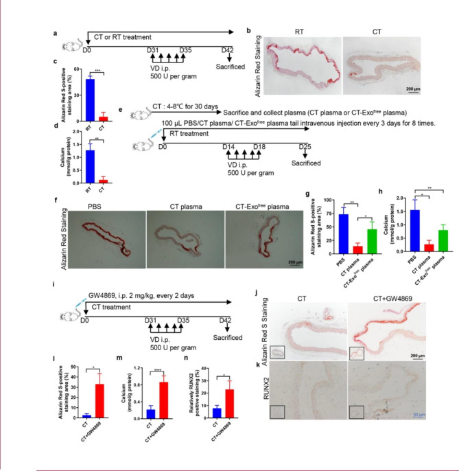
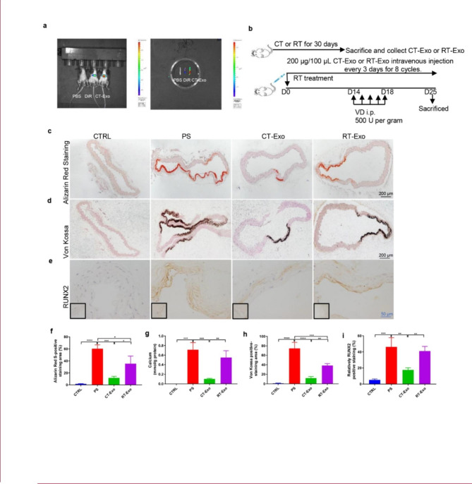
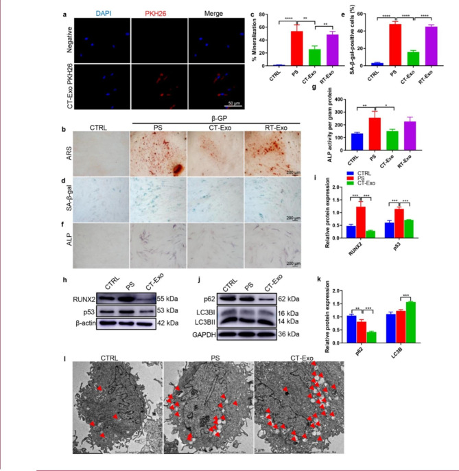
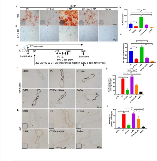
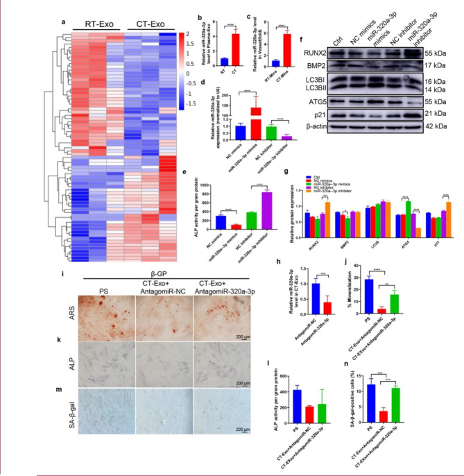
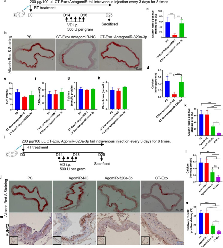
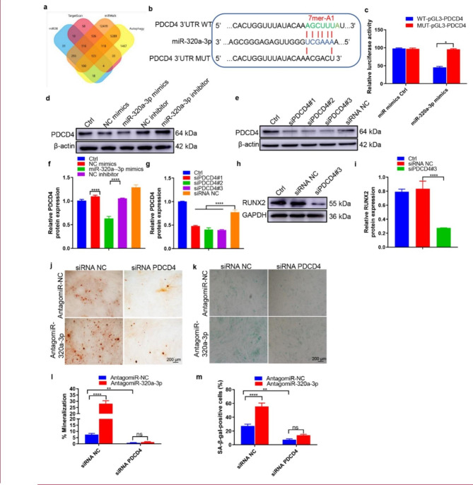
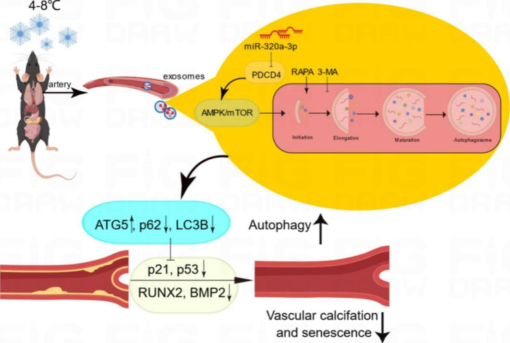
Medial arterial calcification (MAC), a systemic vascular disease different from atherosclerosis, is associated with an increased incidence of cardiovascular events. Several studies have demonstrated that ambient temperature is one of the most important factors affecting cardiovascular events. However, there has been limited research on the effect of different ambient temperatures on MAC. In the present study, we showed that cold temperature exposure (CT) in mice slowed down the formation of vitamin D (VD)-induced vascular calcification compared with room temperature exposure (RT). To investigate the mechanism involved, we isolated plasma-derived exosomes from mice subjected to CT or RT for 30 days (CT-Exo or RT-Exo, respectively). Compared with RT-Exo, CT-Exo remarkably alleviated the calcification/senescence formation of vascular smooth muscle cells (VSMCs) and promoted autophagy by activating the phosphorylation of AMP-activated protein kinase (p-AMPK) and inhibiting phosphorylation of mammalian target of rapamycin (p-mTOR). At the same time, CT-Exo promoted autophagy in β-glycerophosphate (β-GP)-induced VSMCs. The number of autophagosomes and the expression of autophagy-related proteins ATG5 and LC3B increased, while the expression of p62 decreased. Based on a microRNA chip microarray assay and real-time polymerase chain reaction, miR-320a-3p was highly enriched in CT-Exo as well as thoracic aortic vessels in CT mice. miR-320a-3p downregulation in CT-Exo using AntagomiR-320a-3p inhibited autophagy and blunted its anti-calcification protective effect on VSMCs. Moreover, we identified that programmed cell death 4 (PDCD4) is a target of miR-320a-3p, and silencing PDCD4 increased autophagy and decreased calcification in VSMCs. Treatment with CT-Exo alleviated the formation of MAC in VD-treated mice, while these effects were partially reversed by GW4869. Furthermore, the anti-arterial calcification protective effects of CT-Exo were largely abolished by AntagomiR-320a-3p in VD-induced mice. In summary, we have highlighted that prolonged cold may be a good way to reduce the incidence of MAC. Specifically, miR-320a-3p from CT-Exo could protect against the initiation and progression of MAC via the AMPK/mTOR autophagy pathway.

Keywords: Arterial calcification; Autophagy; Cold exposure; PDCD4; Plasma-derived exosomes; Senescence; miR-320a-3p.


Conflict of interest statement

The authors declare no competing interests.

Figures

Fig. 1
Fig. 1
Cold exposure protected against MAC in a VD-induced mouse model. (a) The schematic flow diagram represents the in vivo treatment of CT or RT in the VD-treated mouse model (n = 6 per group). ARS-stained sections from thoracic aorta (b) and quantitation of positive staining area (c) are shown. The black scale bar is 200 μm. (d) Vascular calcium content measurement. (e) Experimental design of the VD-induced vascular calcification mouse model treated with PBS, CT plasma or CT-Exofree plasma by intravenous injection (n = 6 per group). ARS-stained sections from thoracic aorta (f) and quantitation of the positive staining area (g) are shown. The black scale bar is 200 μm. (h) Calcium content of the thoracic aorta. (i) Schematic flow diagram represented the in vivo treatment of CT with or without GW4869 in the VD-induced mice model (n = 6 per group). Evaluation of the effect of pre-treatment of the exosome blocker GW4869 on arterial calcification induced by VD calcified mice in CT treatment. ARS staining (j, l) and RUNX2 expression (k, n) analysis of paraffin-embedded vascular tissue from mice. (m) Vascular calcium content measurement. The black scale bar is 200 μm and the blue scale bar is 50 μm. The data are presented as the mean ± standard deviation with three replicates for each group. The data were analysed with Student’s t-test or one-way ANOVA with the Bonferroni post hoc test. *p < 0.05; **p < 0.01; ***p < 0.001; ****p < 0.0001
Fig. 2
Fig. 2
CT-Exo protected against vascular calcification in the VD-induced mouse model. (a) Uptake of DiR-labelled CT-Exo in aortic VSMCs of mice. The mice were subjected to the intravenous administration of PBS, DiR or DiR-labelled CT-Exo treatments (100 µg/mice, n = 3 per group). Representative in vivo fluorescence image of CT-Exo distribution in mice 24 h after CT-Exo injection. (b) Experimental design of the VD-induced vascular calcification mouse model treated with PBS, CT-Exo or RT-Exo by intravenous injection (n = 6 per group). ARS (c) and Von Kossa staining (d) and quantification of the percentages of ARS+ (f) and Von Kossa+ (h) areas. (g) Vascular calcium content measurement. RUNX2 expression in thoracic aorta (e) and quantitation of positive staining area (i) are shown. The black scale bar is 200 μm and the blue scale bar is 50 μm. The CTRL group represents the negative control group with only PBS treatment. The PS group represents the positive control group with only β-GP treatment. The data are presented as the mean ± standard deviation with three replicates for each group. The data were analysed with one-way ANOVA with the Bonferroni post hoc test or the unpaired, two-tailed Student’s t-test. *p < 0.05; **p < 0.01; ***p < 0.001; ****p < 0.0001
Fig. 3
Fig. 3
CT-Exo protected VSMCs against calcification by promoting autophagy. (a) Representative fluorescence micrograph of PKH26-labelled CT-Exo (red) internalised by VSMCs; nuclei are shown in blue. The white scale bar is 50 μm. ARS (b) and SA-β-gal (d) staining was evaluated in VSMCs incubated with β-GP and CT-Exo for 28 and 10 days, respectively. n = 5, the black scale bar is 200 μm. (c, e) The data are presented as ratio of positive staining area. (f) ALP staining was measured in VSMCs incubated with β-GP and CT-Exo for 14 days. The black scale bar is 200 μm. (g) ALP activity. (h) RUNX2 and p53 protein expression was determined by western blotting after β-GP and CT-Exo treatment for 3 days. The data are presented as densitometric ratios normalised to β-actin (i), n = 4. (j, k) Western blots (j) and quantification (k) of p62 and LC3B in the PBS, PS and CT-Exo VSMCs, n = 4. (l) VSMCs were incubated with β-GP and CT-Exo for 72 h and then analysed by electron microscopy; a representative image is shown. Autophagosomes containing organelle remnants are highlighted by red arrows (n = 4 per group). The PS group represents the control group with only β-GP treatment. Each experiment was repeated three times. The data are presented as the mean ± standard deviation with three replicates. The data were analysed with one-way ANOVA with the Bonferroni post hoc test. *p < 0.05; **p < 0.01; ***p < 0.001; ****p < 0.0001
Fig. 4
Fig. 4
3-MA attenuated the pro-aging/pro-calcification preventive effect of CT-Exo in vitro and in vivo. Representative images of ARS (a) and SA-β-gal (c) staining of VSMCs that had been pre-treated with the indicated concentrations of 3-MA or RAPA for 30 min and then incubated with β-GP for 28 and 10 days, respectively. n = 5, the scale bar is 200 μm. Quantitative analysis of the percentages of ARS+ (b, in red) and SA-β-gal+ (d, in green) areas. (e) Schematic illustration of the experimental design used to assess the effects of CT-Exo and 3-MA on the vascular phenotype in VD-induced mice (n = 6 per group). (f, g) Von Kossa staining showed calcified aorta from CRTL, PS, CT-Exo, 3-MA, CT-Exo + 3-MA and RAPA mice (n = 6 per group). The black scale bar is 200 μm. (h, i) p21 expression in aorta from the six groups of mice were examined by immunohistochemistry. The black scale bar is 200 μm (n = 6 per group). The CTRL group represents the negative control group with only PBS treatment. The PS group represents the positive control group with only β-GP treatment. The data are presented as the mean ± standard deviation. The data were analysed with one-way ANOVA with the Bonferroni post hoc test. *p < 0.05; **p < 0.01; ***p < 0.001; ****p < 0.0001
Fig. 5
Fig. 5
miR-320a-3p antagonised osteogenic differentiation of VSMCs. (a) The heatmap shows the differentially expressed miRNAs (absolute fold change ≥ 1.5, p < 0.05) between CT-Exo and RT-Exo (n = 3 per group). (b) qRT-PCR analysis of miR-320a-3p expression in exosomes from the plasma of the RT or CT mice (n = 6). (c) qRT-PCR analysis of miR-320a-3p expression in vessel s from RT or CT mice (n = 6). (d) qRT-PCR was performed to evaluate the expression of miR-320a-3p in VSMCs transfected with specific miR-320a-3p mimics or inhibitor (n = 4). (e) The ALP activity was evaluated by using specific kits in VSMCs transfected with specific miR-320a-3p mimics or inhibitors (n = 4). (f) Western blotting was performed to determine the protein expression of RUNX2, BMP2, LC3B, ATG5 and p21 in VSMCs transfected with specific miR-320a-3p mimics or inhibitors (n = 4). (g) The data are presented as densitometric ratios normalised to β-actin. (h) qRT-PCR analysis of miR-320a-3p expression in CT-Exo + AntagomiR-320a-3p (n = 6). ARS staining (i, j), ALP staining (k) and ALP activity (l) quantification of SA-β-gal-stained positive cells was shown (m, n). The black scale bar represents 200 μm (n = 5 per group). The PS group represents the positive control group with only β-GP treatment. The data are presented as the mean ± standard deviation. The data were analysed with one-way ANOVA with the Bonferroni post hoc test or the unpaired, two-tailed Student’s t-test. *p < 0.05; **p < 0.01; ***p < 0.001; ****p < 0.0001
Fig. 6
Fig. 6
miR-320a-3p effectively protected against MAC in vivo and its related biochemical indicators. (a) Experimental design of the VD-induced vascular calcification mouse model treated with PBS, CT-Exo + AntagomiR-NC or CT-Exo + AntagomiR-320a-3p by intravenous injection (n = 6 per group). ARS staining and quantitation (b, c) and vascular calcium content measurement (d). The black scale bar is 200 μm. Serum BUN (e), CREA (f), calcium (g) and phosphate (h) levels in mice with VD-induced vascular calcification (n = 6). (i) Experimental design of the VD-induced vascular calcification mouse model treated with PBS, AgomiR-NC or AgomiR-320a-3p by intravenous injection (n = 6). ARS staining and quantitation (j, k) and vascular calcium content measurement (l). RUNX2 expression in the thoracic aorta (m) and quantitation of positive staining area (n) are shown. The black scale bar is 200 μm and the blue scale bar is 50 μm. The PS group represents the control group with only β-GP treatment. The data are presented as the mean ± standard deviation. The data were analysed with one-way ANOVA with the Bonferroni post hoc test. ns > 0.05; *p < 0.05; **p < 0.01; ***p < 0.001 and ****p < 0.0001
Fig. 7
Fig. 7
PDCD4 was a direct target gene of miR-320a-3p and regulated VSMCs calcification. (a) A Venn diagram showing bioinformatics analysis of miR-320a-3p target genes. (b) Schematic representation of miR-320a-3p putative target sites in the PDCD4 3′-UTR and the alignment of miR-320a-3p with wild type and mutant PDCD4 3′-UTR showing pairing. (c) Luciferase reporter assays were performed using luciferase constructs carrying a wild type or mutant PDCD4 3′-UTR co-transfected into VSMCs with miR-320a-3p mimics compared with empty vector control. Firefly luciferase activity was normalised to Renilla luciferase activity. (d, f) PDCD4 protein expression in VSMCs transfected with miR-320a-3p mimics or miR-320a-3p inhibitor was determined by western blot (n = 4). (e and g) The efficiency of PDCD4 knockdown in VSMCs by siRNA was measured by western blot (n = 4). (h-i) RUNX2 expression was measured in the VSMCs treated with siPDCD4#3 or siRNA control (n = 4). (j) ARS staining in β-GP-treated VSMCs transfected with inhibitors of miR-320a-3p in the presence or absence of PDCD4 siRNA for 28 days; representative micrographs are shown. (K) SA-β-gal staining was measured in VSMCs incubated with β-GP for 10 days. n = 4, the data are presented as the ratio of positive ARS (j) and SA-β-gal (m) staining area. The scale bar is 200 μm. The data are presented as the mean ± standard deviation. The data were analysed with one or two-way ANOVA with the Bonferroni post hoc test. ns > 0.05; *p < 0.05; **p < 0.01; ***p < 0.001 and ****p < 0.0001
Fig. 8
Fig. 8
CT-Exo enrichment of miR-320a-3p under CT exposure can protect against vascular calcification and senescence by activating autophagy through the AMPK/mTOR pathway. CT-Exo with the high expression of miR-320a-3p can be secreted from mice plasma exposed to a cold environment. PDCD4 was found to be a potential target of miR-320a-3p and to increase osteogenic differentiation and senescence of VSMCs. CT-Exo can activate AMPK/mTOR, a classical autophagy pathway and then activate the expression of autophagy proteins (LC3B and ATG5) and reduce the degradation of autophagy specific substrates (p62). Ultimately, this slow down the level of senescence (p21 and p53) and decrease the level of calcification (RUNX2 and BMP2) of VSMCs

References

    1. Wee NKY, Nguyen AD, Enriquez RF, Zhang L, Herzog H, Baldock PA. Neuropeptide Y Regulation of Energy Partitioning and Bone Mass during Cold exposure. Calcif Tissue Int. 2020;107(5):510–23. - PubMed
    1. Dong M, Yang X, Lim S, Cao Z, Honek J, Lu H, Zhang C, Seki T, Hosaka K, Wahlberg E, Yang J, Zhang L, Länne T, Sun B, Li X, Liu Y, Zhang Y, Cao Y. Cold exposure promotes atherosclerotic plaque growth and instability via UCP1-dependent lipolysis. Cell Metab. 2013;18(1):118–29. - PMC - PubMed
    1. Sacks D, Baxter B, Campbell BCV, Carpenter JS, Cognard C, Dippel D, Eesa M, Fischer U, Hausegger K, Hirsch JA, Shazam Hussain M, Jansen O, Jayaraman MV, Khalessi AA, Kluck BW, Lavine S, Meyers PM, Ramee S, Rüfenacht DA, Schirmer CM, Vorwerk D. Multisociety Consensus Quality Improvement revised Consensus Statement for Endovascular Therapy of Acute ischemic stroke. Int J Stroke. 2018;13(6):612–32. - PubMed
    1. Du J, He Z, Xu M, Qu X, Cui J, Zhang S, Zhang S, Li H, Yu Z. Brown Adipose tissue rescues bone loss Induced by Cold exposure. Front Endocrinol (Lausanne) 2021;12:778019. - PMC - PubMed
    1. Serrat MA. Environmental temperature impact on bone and cartilage growth. Compr Physiol. 2014;4(2):621–55. - PubMed