. 2023 Jul 17;22(1):113.
doi: 10.1186/s12943-023-01804-z.

Exosome-mediated miR-144-3p promotes ferroptosis to inhibit osteosarcoma proliferation, migration, and invasion through regulating ZEB1

Affiliations

Exosome-mediated miR-144-3p promotes ferroptosis to inhibit osteosarcoma proliferation, migration, and invasion through regulating ZEB1

Mingyang Jiang et al. Mol Cancer. .

Abstract

Background: Osteosarcoma (OS) is the most prevalent orthopedic malignancy with a dismal prognosis. The high iron absorption rate in OS cells of patients suggests that ferroptosis may be related to the progression of OS, but its potential molecular regulatory role is still unclear. Based on the ability to couple with exosomes for targeted delivery of signals, exosome-derived micro ribonucleic acids (miRNAs) can potentially serve as diagnostic biomarkers for OS.

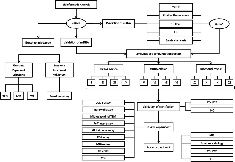
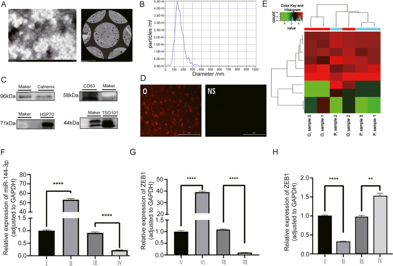
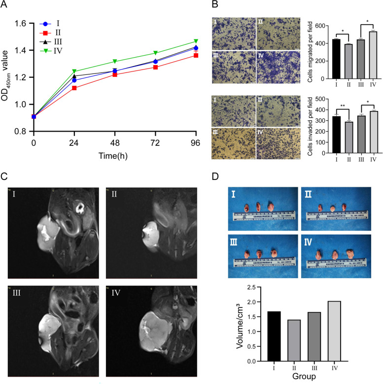
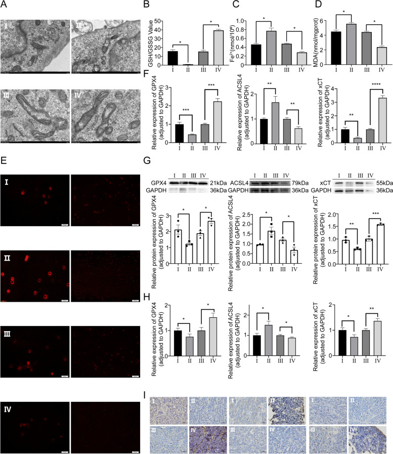
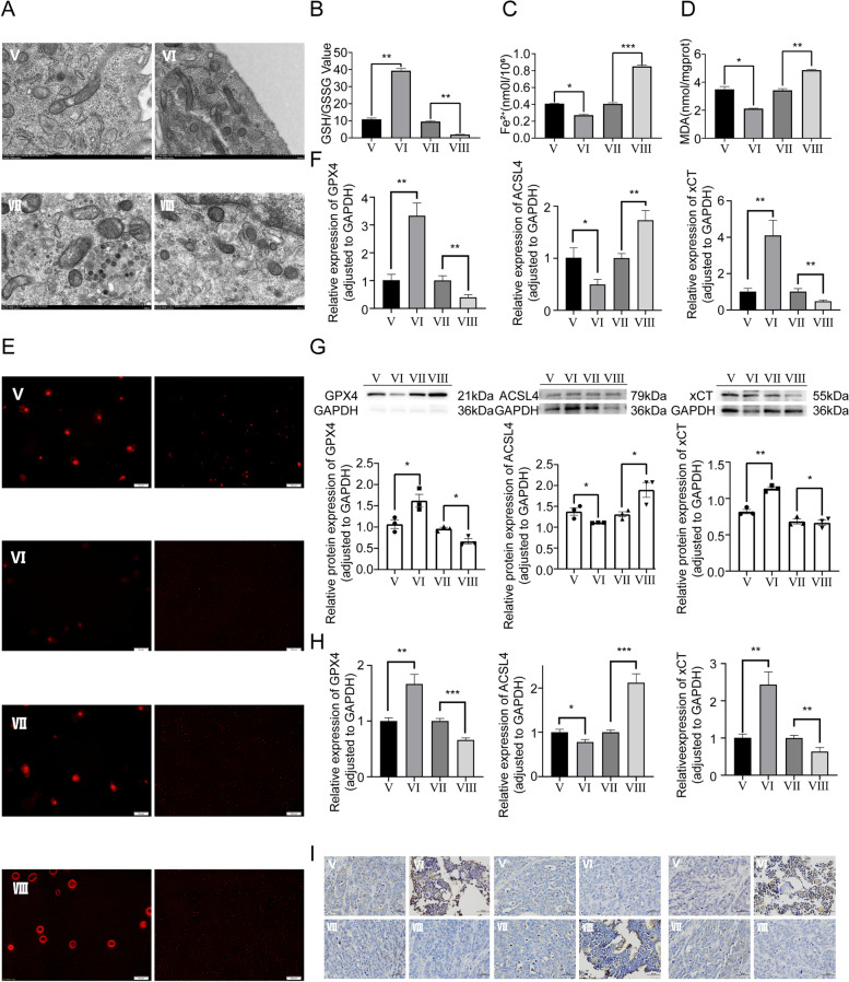
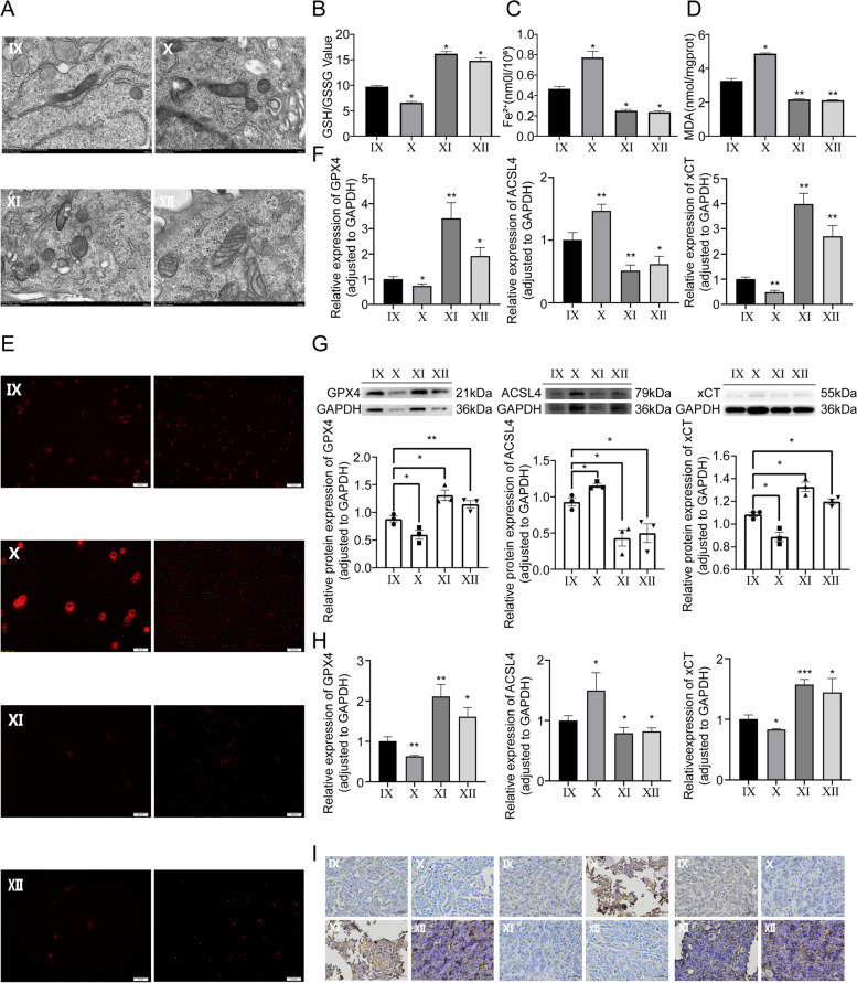
Methods: We identified ferroptosis-related miRNAs and messenger ribonucleic acids(mRNAs) in OS using bioinformatics analysis and performed survival analysis. Then we measured miRNA expression levels through exosome microarray sequencing, and used RT-qPCR and IHC to verify the expression level of miR-144-3p and ZEB1. Stable gene expression cell lines were fabricated for in vitro experiments. Cell viability, migration and invasion were determined by CCK-8 and transwell experiment. Use the corresponding reagent kit to detect GSH/GSSG ratio, Fe2+ level, MDA level and ROS level, and measure the expression levels of GPX4, ACSL4 and xCT through RT-qPCR and WB. We also constructed nude mice model for in vivo experiments. Finally, the stability of the miRNA/mRNA axis was verified through functional rescue experiments.

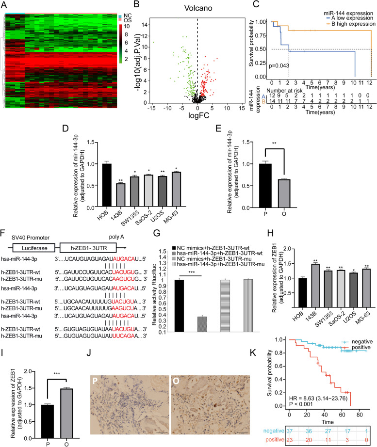
Results: Low expression of miR-144-3p and high expression of ZEB1 in OS cell lines and tissues was observed. Overexpression of miR-144-3p can promote ferroptosis, reduce the survival ability of OS cells, and prevent the progression of OS. In addition, overexpression of miR-144-3p can downregulate the expression of ZEB1 in cell lines and nude mice. Knockdown of miR-144-3p has the opposite effect. The functional rescue experiment validated that miR-144-3p can regulate downstream ZEB1, and participates in the occurrence and development of OS by interfering with redox homeostasis and iron metabolism.

Conclusions: MiR-144-3p can induce the occurrence of ferroptosis by negatively regulating the expression of ZEB1, thereby inhibiting the proliferation, migration, and invasion of OS cells.

Keywords: Exosome; Ferroptosis; Osteosarcoma; ZEB1; miR-144-3p.


Conflict of interest statement

The authors declare no competing interests.

Figures

Fig. 1
Fig. 1
Graphical abstract. Groups of miR-144-3p/ZEB1 edition and functional rescue experiment. I: Normal control group of overexpress miR-144-3p. II: Overexpress miR-144-3p group. III: Normal control group of knockdown miR-144-3p. IV: Knockdown miR-144-3p group. V: Normal control group of overexpression ZEB1. VI: Overexpress ZEB1 group. VII: Normal control group of knockdown ZEB1. VIII: Knockdown ZEB1 group. IX: Normal control group of overexpress miR-144-3p & normal control group of overexpress ZEB1 group. X: Overexpress miR-144-3p & normal control group of overexpress ZEB1 group. XI: Normal control group of overexpress miR-144-3p & overexpress ZEB1 group. XII: Overexpress miR-144-3p & overexpress ZEB1 group
Fig. 2
Fig. 2
Identification and validation of key miRNA and downstream mRNA. A The heatmap and B volcano plot showing the profile of differentially expressed genes based on dataset GSE28425 in the context of OS. C The Kaplan–Meier plot related miR-144-3p expression based on dataset GSE39052. The RT-qPCR results of key miRNA based on D five OS cell lines and one osteoblastic cell line, as well as E human OS tissue and paracancerous tissue samples. F The binding site sequence of miR-144-3p and ZEB1. G The dual luciferase assay indicated that miR-144-3p can bind to ZEB1. The RT-qPCR results of key mRNA based on H five OS cell lines and one osteoblastic cell line, as well as I human OS tissue and paracancerous tissue samples. J IHC micrographs based on 60 tissue sections. K The Kaplan–Meier plot for ZEB1 based on the clinical data of 60 patients corresponding to tissue sections
Fig. 3
Fig. 3
The validation of exosome expression, function and downstream miR-144-3p/ZEB1 axis. A TEM micrographs of exosomes. B The NTA indicated the particle size, volume, and concentration of exosomes. C The WB of exosomes characteristic proteins. D The co-culture assay indicated that labeled exosomes can enter OS cells. E The heatmap of exosome microarray. The RT-qPCR results for validation of transfection F miR-144-3p in Group I-IV and G ZEB1 in Group V-VIII. The RT-qPCR results for validation of downstream H ZEB1 in Groups I-IV
Fig. 4
Fig. 4
Effect of miR-144-3p expression on OS proliferation, invasion and migration. A CCK-8 assay. B Transwell assay of migration and invasion. C MRI images. D Gross samples
Fig. 5
Fig. 5
Effect of miR-144-3p expression on ferroptosis mechanism. A TEM micrographs of mitochondria. B Glutathione assay. C Fe2+ level assay. D MDA assay. E TEM micrographs of immunofluorescence for ROS. F The RT-qPCR and G WB assay of GPX4, ACSL4, and xCT in vitro experiment. H The RT-qPCR and I IHC assay of GPX4, ACSL4, and xCT in vivo experiment
Fig. 6
Fig. 6
Effect of ZEB1 expression on OS proliferation, invasion and migration. A CCK-8 assay. B Transwell assay of migration and invasion. C MRI images. D Gross samples
Fig. 7
Fig. 7
Effect of ZEB1 expression on ferroptosis mechanism. A TEM micrographs of mitochondria. B Glutathione assay. C Fe2+ level assay. D MDA assay. E TEM micrographs of immunofluorescence for ROS. F The RT-qPCR and G WB assay of GPX4, ACSL4, and xCT in vitro experiment. H The RT-qPCR and (I) IHC assay of GPX4, ACSL4, and xCT in vivo experiment
Fig. 8
Fig. 8
Effect of miR-144-3p/ZEB1 axis on OS proliferation, invasion and migration. A CCK-8 assay. B Transwell assay of migration and invasion. C MRI images. D Gross samples
Fig. 9
Fig. 9
Effect of miR-144-3p/ZEB1 axis on ferroptosis mechanism. A TEM micrographs of mitochondria. B Glutathione assay. C Fe2+ level assay. D MDA assay. E TEM micrographs of immunofluorescence for ROS. F The RT-qPCR and G WB assay of GPX4, ACSL4, and xCT in vitro experiment. H The RT-qPCR and I IHC assay of GPX4, ACSL4, and xCT in vivo experiment

References

    1. Bielack SS, Kempf-Bielack B, Delling Gn Gn, Exner GU, Flege S, Helmke K, Kotz R, Salzer-Kuntschik M, Werner M, Winkelmann W, et al. Prognostic factors in high-grade osteosarcoma of the extremities or trunk: an analysis of 1,702 patients treated on neoadjuvant Cooperative Osteosarcoma Study Group protocols. J Clin Oncol. 2002;20:776–90. doi: 10.1200/JCO.2002.20.3.776. - DOI - PubMed
    1. Faisham WI, Mat Saad AZ, Alsaigh LN, Nor Azman MZ, Kamarul Imran M, Biswal BM, Bhavaraju VM, Salzihan MS, Hasnan J, Ezane AM, et al. Prognostic factors and survival rate of osteosarcoma: a single-institution study. Asia Pac J Clin Oncol. 2017;13:e104–e110. doi: 10.1111/ajco.12346. - DOI - PubMed
    1. Anderson ME. Update on survival in osteosarcoma. Orthop Clin North Am. 2016;47:283–292. doi: 10.1016/j.ocl.2015.08.022. - DOI - PubMed
    1. Li Z, Li X, Xu D, Chen X, Li S, Zhang L, Chan MTV, Wu WKK. An update on the roles of circular RNAs in osteosarcoma. Cell Prolif. 2021;54:e12936. doi: 10.1111/cpr.12936. - DOI - PMC - PubMed
    1. Tang F, Min L, Seebacher NA, Li X, Zhou Y, Hornicek FJ, Wei Y, Tu C, Duan Z. Targeting mutant TP53 as a potential therapeutic strategy for the treatment of osteosarcoma. J Orthop Res. 2019;37:789–798. doi: 10.1002/jor.24227. - DOI - PubMed

Publication types