. 2023 Jul 17;21(1):473.
doi: 10.1186/s12967-023-04343-9.

IL-1RA promotes oral squamous cell carcinoma malignancy through mitochondrial metabolism-mediated EGFR/JNK/SOX2 pathway

Affiliations

IL-1RA promotes oral squamous cell carcinoma malignancy through mitochondrial metabolism-mediated EGFR/JNK/SOX2 pathway

Shyng-Shiou F Yuan et al. J Transl Med. .

Abstract

Background: Interleukin-1 receptor antagonist (IL-1RA), a member of the IL-1 family, has diverse roles in cancer development. However, the role of IL-1RA in oral squamous cell carcinoma (OSCC), in particular the underlying mechanisms, remains to be elucidated.

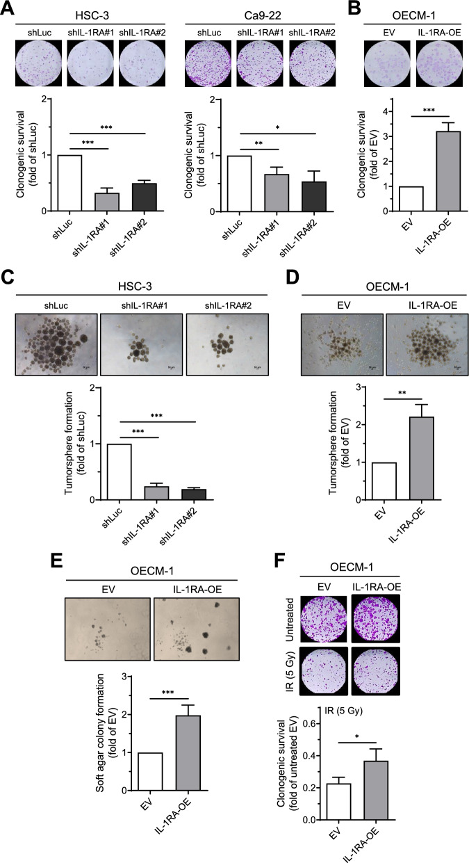
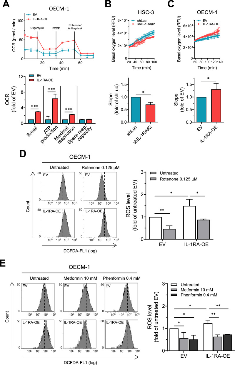
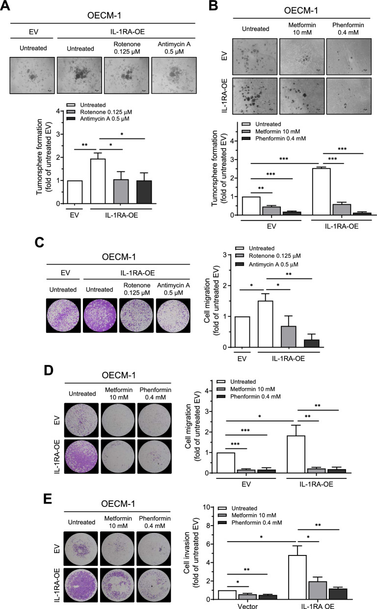
Methods: Tumor tissues from OSCC patients were assessed for protein expression by immunohistochemistry. Patient survival was evaluated by Kaplan-Meier curve analysis. Impact of differential IL-1RA expression on cultured OSCC cell lines was assessed in vitro by clonogenic survival, tumorsphere formation, soft agar colony formation, and transwell cell migration and invasion assays. Oxygen consumption rate was measured by Seahorse analyzer or multi-mode plate reader. PCR array was applied to screen human cancer stem cell-related genes, proteome array for phosphorylation status of kinases, and Western blot for protein expression in cultured cells. In vivo tumor growth was investigated by orthotopic xenograft in mice, and protein expression in xenograft tumors assessed by immunohistochemistry.

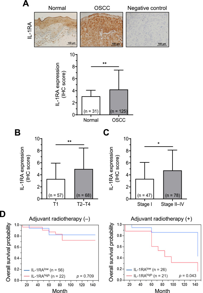
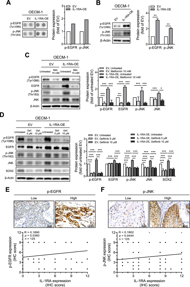
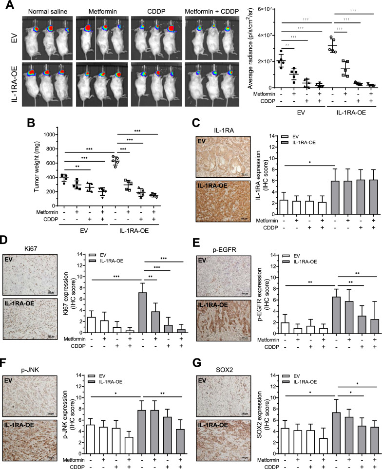
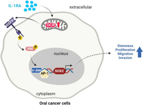
Results: Clinical analysis revealed that elevated IL-1RA expression in OSCC tumor tissues was associated with increased tumor size and cancer stage, and reduced survival in the patient group receiving adjuvant radiotherapy compared to the patient group without adjuvant radiotherapy. In vitro data supported these observations, showing that overexpression of IL-1RA increased OSCC cell growth, migration/invasion abilities, and resistance to ionizing radiation, whereas knockdown of IL-1RA had largely the opposite effects. Additionally, we identified that EGFR/JNK activation and SOX2 expression were modulated by differential IL-1RA expression downstream of mitochondrial metabolism, with application of mitochondrial complex inhibitors suppressing these pathways. Furthermore, in vivo data revealed that treatment with cisplatin or metformin-a mitochondrial complex inhibitor and conventional therapy for type 2 diabetes-reduced IL-1RA-associated xenograft tumor growth as well as EGFR/JNK activation and SOX2 expression. This inhibitory effect was further augmented by combination treatment with cisplatin and metformin.

Conclusions: The current study suggests that IL-1RA promoted OSCC malignancy through mitochondrial metabolism-mediated EGFR/JNK activation and SOX2 expression. Inhibition of this mitochondrial metabolic pathway may present a potential therapeutic strategy in OSCC.

Keywords: Cancer stemness; EGFR; IL-1RA; JNK; Metastasis; Mitochondrial metabolism; OSCC; SOX2; Tumor growth.


Conflict of interest statement

The authors declare that they have no competing interests.

Figures

Fig. 1
Fig. 1
Association of IL-1RA expression with OSCC progression and patient outcomes. A Protein expression of IL-1RA in oral tumor tissues from OSCC patients (OSCC) and normal oral tissues from normal epithelial regions of fibroma patients (Normal) was analyzed by immunohistochemistry (IHC). The negative control for IL-1RA staining was performed in parallel with omission of primary antibodies. BC Protein expression of IL-1RA in OSCC tumor tissues was analyzed by IHC for the comparison of primary tumor size (T1 versus T2-T4) in (B), and cancer stage (Stage I versus Stage II-IV) in (C). The scoring of IHC was described in the Methods section. Data were presented as mean ± SD with the indicated sample size in parentheses. *, p < 0.05; **, p < 0.01. D Overall survival probability was analyzed by Kaplan–Meier survival curves for patients with adjuvant radiotherapy ( +) and patients without adjuvant radiotherapy (−) according to the expression level of IL-1RA in OSCC tumor tissues (low versus high) as described in the Methods section. The sample size in each group was indicated in parentheses
Fig. 2
Fig. 2
Effect of IL-1RA expression on in vitro cell growth of OSCC cells. AF Following knockdown of IL-1RA expression in HSC-3 and Ca9-22 cells and overexpression of IL-1RA in OECM-1 cells, assessments were performed for clonogenic survival (A, B), tumorsphere formation (C, D), and colony formation in soft agar (E). In (F), clonogenic survival was assessed in OECM-1 cells with overexpression of IL-1RA and control cells in the presence or absence of ionizing radiation treatment. Data were presented as mean ± SD from three independent experiments. *, p < 0.05; **, p < 0.01; ***, p < 0.001. shLuc, knockdown of firefly luciferase; shIL-1RA, knockdown of IL-1RA; EV, empty vector; IL-1RA-OE, overexpression of IL-1RA
Fig. 3
Fig. 3
Effect of IL-1RA expression on mitochondrial metabolism and reactive oxygen species (ROS) formation in OSCC cells. A Multiple status of mitochondrial metabolism was measured by oxygen consumption rate (OCR) using Agilent Seahorse XFe24 Analyzer via sequential delivery of the indicated mitochondrial modulators (oligomycin, FCCP, and a mixture of rotenone and antimycin A) to OECM-1 cells with overexpression of IL-1RA and control cells. BC Basal OCR indicated by the slope (∆RFU/∆min) was measured with CLARIOstar Plus multi-mode plate reader using a commercial oxygen consumption-labeling kit, for HSC-3 cells with knockdown of IL-1RA and control cells in (B), and OECM-1 cells with overexpression of IL-1RA and control cells in (C). DE ROS formation was measured by flow cytometry after DCFDA labeling of OECM-1 cells with overexpression of IL-1RA and control cells in the presence or absence of rotenone in (D), and metformin or phenformin in (E). Data were presented as mean ± SD from three independent experiments. *, p < 0.05; **, p < 0.01; ***, p < 0.001. RFU, relative fluorescence unit; shLuc, knockdown of firefly luciferase; shIL-1RA, knockdown of IL-1RA; EV, empty vector; IL-1RA-OE, overexpression of IL-1RA
Fig. 4
Fig. 4
Involvement of mitochondrial metabolism in IL-1RA-promoted malignant behaviors of OSCC cells. AB OECM-1 cells with IL-1RA overexpression and control cells were assessed for tumorsphere formation in the presence or absence of rotenone or antimycin A in (A), and metformin or phenformin in (B). CD OECM-1 cells with IL-1RA overexpression and control cells were assessed for transwell cell migration ability in the presence or absence of rotenone or antimycin A in (C), and metformin or phenformin in (D). E OECM-1 cells with IL-1RA overexpression and control cells were assessed for transwell cell invasion ability in the presence or absence of metformin or phenformin. Data were presented as mean ± SD from three independent experiments. *, p < 0.05; **, p < 0.01; ***, p < 0.001. EV, empty vector; IL-1RA-OE, overexpression of IL-1RA
Fig. 5
Fig. 5
Involvement of mitochondrial metabolism in the expression of IL-1RA-associated cancer stemness markers. A PCR array was applied to screen a set of cancer stem cell-related genes in HSC-3 cells with knockdown of IL-1RA, in which the expression of each gene was quantitated relative to that in control cells (dashed line). BC Four candidates (SOX2, THY1, EGF, and KIT), as indicated by the solid rectangle in (A), were further examined by Western blot for their protein expression in HSC-3 and Ca9-22 cells with knockdown of IL-1RA in (B), and OECM-1 cells with overexpression of IL-1RA in (C), along with the control cells. D Protein expression of SOX2 in OECM-1 cells with overexpression of IL-1RA and control cells was examined by Western blot in the presence or absence of rotenone or antimycin A. Data were presented as mean ± SD from three independent experiments in (BD). *, p < 0.05; **, p < 0.01; ***, p < 0.001. Rot., rotenone; Ant. A, antimycin A; shLuc, knockdown of firefly luciferase; shIL-1RA, knockdown of IL-1RA; EV, empty vector; IL-1RA-OE, overexpression of IL-1RA
Fig. 6
Fig. 6
Involvement of mitochondrial metabolism in IL-1RA-associated EGFR/JNK activation and SOX2 expression. AB Up-regulated phosphorylation of EGFR and JNK was shown by phospho-kinase proteome array screening for the phosphorylation status of a set of kinases, with duplicate detection for each kinase, in OECM-1 cells with overexpression of IL-1RA in (A), which was further confirmed by Western blot in (B). C Protein expression of phosphorylated EGFR and JNK, along with their unphosphorylated forms, in OECM-1 cells with overexpression of IL-1RA and control cells was examined by Western blot in the presence or absence of metformin. D Protein expression of phosphorylated EGFR and JNK, along with their unphosphorylated forms, and SOX2 in OECM-1 cells with overexpression of IL-1RA and control cells was examined by Western blot in the presence or absence of gefitinib. Data were presented as mean ± SD from three independent experiments in (BD). *, p < 0.05; **, p < 0.01; ***, p < 0.001. EF Correlation between IL-1RA expression and phosphorylation of EGFR (E) and JNK (F) in OSCC tumor tissues from patients was analyzed according to IHC scoring, and was evaluated by Pearson correlation (r). Met., metformin; Gef., gefitinib; p-EGFR, phosphorylated EGFR; p-JNK, phosphorylated JNK; EV, empty vector; IL-1RA-OE, overexpression of IL-1RA
Fig. 7
Fig. 7
Effect of IL-1RA expression on in vivo tumor growth of OSCC in xenograft mice and the therapeutic potential of metformin. A Orthotopic xenograft of OECM-1 cells with overexpression of IL-1RA or control cells in mice was measured weekly by in vivo imaging system (IVIS) for the groups treated with or without metformin and/or CDDP. The data shown in (A) were the measurements at endpoint of the experiment before sacrifice of the mice. B After sacrifice of the mice at endpoint of the experiment, the xenograft tumors were collected and tumor weight was measured. CG After sacrifice of the mice at endpoint of the experiment, the xenograft tumors were collected and analyzed for protein expression of IL-1RA (C), Ki67 (D), phosphorylated EGFR (E), phosphorylated JNK (F), and SOX2 (G). Data were presented as mean ± SD from five mice in each group. *, p < 0.05; **, p < 0.01; ***, p < 0.001. CDDP, cisplatin; p-EGFR, phosphorylated EGFR; p-JNK, phosphorylated JNK; EV, empty vector; IL-1RA-OE, overexpression of IL-1RA
Fig. 8
Fig. 8
Schematic summary for the current study. The results of the current study suggest that mitochondrial metabolism may be crucial in mediating the IL-1RA-promoted malignant behaviors of OSCC cells, such as cancer stemness-associated tumor growth and migration/invasion abilities, through a pathway involving EGFR/JNK activation and SOX2 expression

References

    1. Du M, Nair R, Jamieson L, Liu Z, Bi P. Incidence trends of lip, oral cavity, and pharyngeal cancers: global burden of disease 1990–2017. J Dent Res. 2020;99(2):143–151. doi: 10.1177/0022034519894963. - DOI - PubMed
    1. Rivera C. Essentials of oral cancer. Int J Clin Exp Pathol. 2015;8(9):11884–11894. - PMC - PubMed
    1. Sung H, Ferlay J, Siegel RL, Laversanne M, Soerjomataram I, Jemal A, Bray F. Global cancer statistics 2020: GLOBOCAN estimates of incidence and mortality worldwide for 36 cancers in 185 countries. CA Cancer J Clin. 2021;71(3):209–249. doi: 10.3322/caac.21660. - DOI - PubMed
    1. Coletta RD, Yeudall WA, Salo T. Grand challenges in oral cancers. Front Oral Health. 2020;1:3. doi: 10.3389/froh.2020.00003. - DOI - PMC - PubMed
    1. Cheng Y, Li S, Gao L, Zhi K, Ren W. The molecular basis and therapeutic aspects of cisplatin resistance in oral squamous cell carcinoma. Front Oncol. 2021;11:761379. doi: 10.3389/fonc.2021.761379. - DOI - PMC - PubMed

Publication types