. 2022 Dec 15:2:98.
doi: 10.1038/s43586-022-00179-7.

Engineered hydrogels for mechanobiology

Affiliations

Engineered hydrogels for mechanobiology

Ulrich Blache et al. Nat Rev Methods Primers. .

Abstract

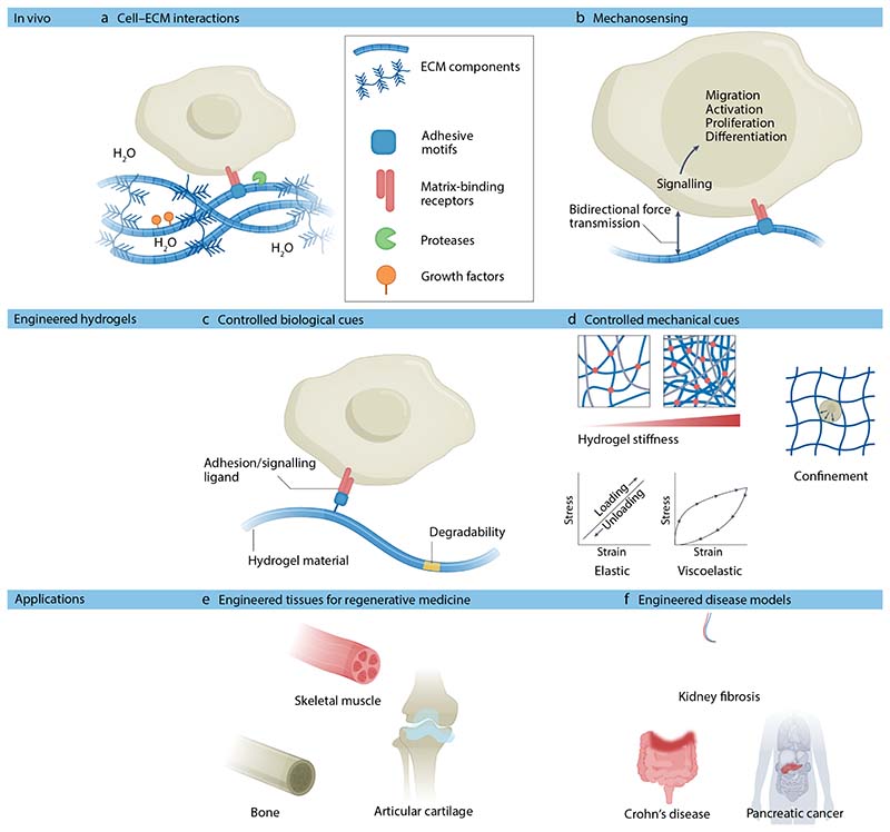
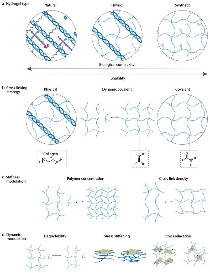
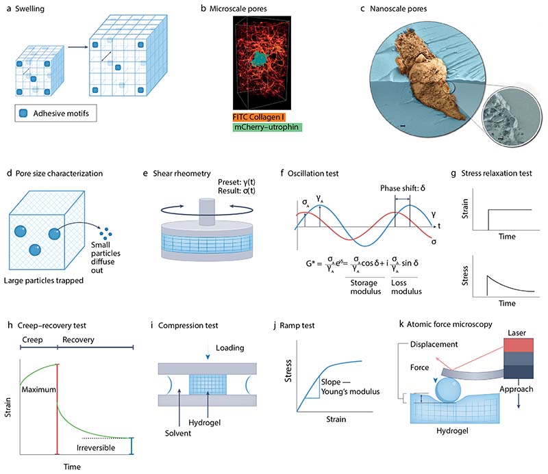
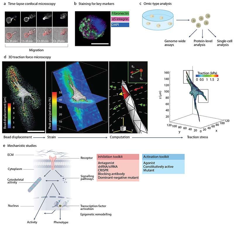
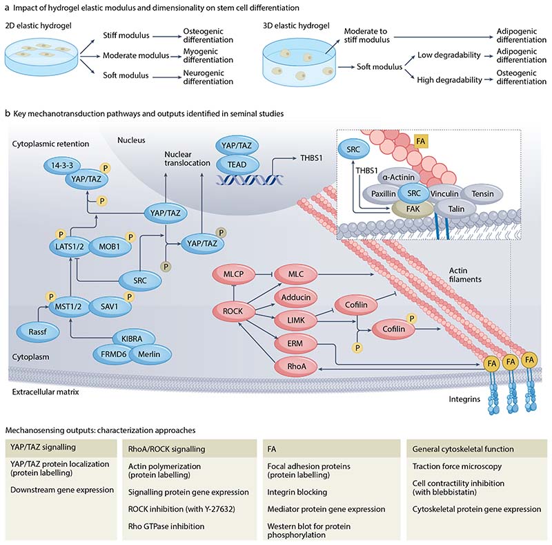
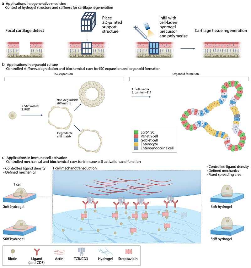
Cells' local mechanical environment can be as important in guiding cellular responses as many well-characterized biochemical cues. Hydrogels that mimic the native extracellular matrix can provide these mechanical cues to encapsulated cells, allowing for the study of their impact on cellular behaviours. Moreover, by harnessing cellular responses to mechanical cues, hydrogels can be used to create tissues in vitro for regenerative medicine applications and for disease modelling. This Primer outlines the importance and challenges of creating hydrogels that mimic the mechanical and biological properties of the native extracellular matrix. The design of hydrogels for mechanobiology studies is discussed, including appropriate choice of cross-linking chemistry and strategies to tailor hydrogel mechanical cues. Techniques for characterizing hydrogels are explained, highlighting methods used to analyze cell behaviour. Example applications for studying fundamental mechanobiological processes and regenerative therapies are provided, along with a discussion of the limitations of hydrogels as mimetics of the native extracellular matrix. The article ends with an outlook for the field, focusing on emerging technologies that will enable new insights into mechanobiology and its role in tissue homeostasis and disease.


Conflict of interest statement

Competing interests The authors declare no competing interests.

Figures

Figure 1
Figure 1. Engineered hydrogels replicate ECM and mechanical cues of native tissues.
a) In native tissues, cells interact with their surrounding ECM via matrix-binding receptors. b) Cells detect mechanical cues in their local environment by applying traction on their surrounding matrix. This process, known as mechanosensing, prompts biochemical signaling, which drives gene transcription and activation of various proteins resulting in cell migration, proliferation and progenitor cell differentiation. c) In vivo-like ECM cues can be replicated in hydrogels, often by tethering adhesive motifs copied from the native ECM directly into the hydrogel material. d) Many mechanical cues provided by the native environment can be replicated using hydrogels. For example, hydrogel properties can be modulated to mimic the stiffness or viscoelastic properties of a native tissue. Minimally deformable and non-degradable matrices can also be created to confine cells. e) Engineered hydrogels have found numerous applications in regenerative medicine and disease modelling. For example, as scaffolds for muscle, bone and cartilage tissue engineering. f) Hydrogels have also been applied in disease modelling to provide a local environment to cells and organoids that mimics tissue-specific ECM ligands and local elasticity/viscoelasticity. This approach enables mimicking of pathological changes in the native ECM during fibrotic wound healing and in diseases such as cancer. Abbreviations: ECM, extracellular matrix.
Figure 2
Figure 2. Overview of different types of hydrogels.
(A) Hydrogels can be formed from natural or synthetic materials or by making hybrid designs that contain both material types. Natural materials show a high degree of bioactivity, but it is often not possible to orthogonally tune their mechanical or dynamic properties. This contrasts with synthetic materials, which are often highly tunable but are limited in their biological complexity. (B) Hydrogels can be formed using covalent, physical or dynamic-covalent cross-linking strategies. (C) Hydrogels’ mechanical properties can be modulated by varying polymer concentration and/or the cross-linking density. (D) Time-dependent properties including stress stiffening, matrix degradability and stress relaxation can be incorporated into hydrogel designs.
Figure 3
Figure 3. Measuring physical properties of hydrogels.
a) Hydrogel swelling and associated changes in mesh size and ligand density. b) Fluorescence light-sheet micrograph showing a neutrophil-like human HL-60 cell expressing mCherry-utrophin in a fluorescently labelled collagen matrix. c) False-coloured cryogenic-scanning electron microscopy (SEM) micrograph showing the 3D interface between a fully hydrated hydrogel (blue) and an encapsulated cell (brown). Scale bar, 1 µm. d) Mesh size characterization using beads with known sizes. Small beads will diffuse within the hydrogel and be detected within the surrounding fluid whereas large beads will remain entrapped, providing an indication of mesh size. e) Operation of rotational rheometer. f) Example of a sinusoidal stress (σ)-strain (γ) curve and how storage and loss moduli are defined using the peak amplitudes (σA,γA) and phase shift (δ). g) General features of material response in a stress relaxation test. h) General features of material response in a creep-recovery test. i) Operation of a compression testing device. j) General behaviour of a hydrogel during a ramp test. k) Operation of an atomic force microscope and general features of a hydrogel response during a cycle of approach and retraction of the atomic force microscope cantilever. Part b reprinted with permission from ref. Part c adapted with permission from ref. Part h reprinted with permission from ref.
Figure 4
Figure 4. Analysis of cells in hydrogels.
a) Time series of images of a cancer cell migrating in a hydrogel using time-lapse confocal microscopy. Top row shows images of RFP-LifeAct whereas bottom row combines brightfield and actin imaging. b, Human mesenchymal stem cell (hMSC) encapsulated in a hydrogel. Immunostaining for fibronectin, α5 integrin, and DAPI. Scale bar = 10µm. c) Omics-type analyses can be used to assess gene expression, chromatin accessibility, protein levels and phosphorylation, and single-cell characteristics. d) Pipeline for determining traction forces generated by cells within a hydrogel from bead displacements. Scale bar = 50µm. e) Mechanotransduction pathways typically involve activation of a membrane receptor, which causes subsequent cytoskeletal activity and signaling pathway activation, impacting epigenetic remodelling and transcription factor activation, together regulating cell activity and phenotype. Specific molecules can be inhibited or activated to assess their role in cell behaviour. Part a is adapted from ref, CC BY 4.0 (https://creativecommons.org/licenses/by/4.0/). Part b is adapted from ref, CC BY 4.0 (https://creativecommons.org/licenses/by/4.0/). Part c is adapted from ref. Part d is adapted from ref, Springer Nature Limited.
Figure 5
Figure 5. Hydrogels for directing stem cell fate by engaging mechanotransduction pathways.
a) Hydrogel modulus and dimensionality impact cellular mechanotransduction. Their impact on promoting human mesenchymal stem cell (hMSC) differentiation is highlighted. Hydrogels can be designed with specific mechanical properties to promote stem cell fate specification. b) Seminal studies with hMSCs, amongst other cell types, have elucidated that mechanotransduction is mediated, in part, by YAP/TAZ and RhoA/ROCK signaling, focal adhesion formation and more generally the cytoskeleton. Key pathways are noted along with approaches for characterizing them. 14-3-3, tyrosine 3-monooxygenase/tryptophan 5-monooxygenase activation protein; ERM, ezrin/radixin/moesin protein family; FA, focal adhesion complex; FAK, focal adhesion kinase; FRMD6, FERM (4.1-ezrin-radixin-moesin) domain-containing protein 6; KIBRA, kidney and brain expressed protein; LATS1/2, serine/threonine-protein kinase 1/2; LIMK, LIM domain kinase; MLC, myosin light chain; MLCP, myosin light chain phosphatase; MOB1, monopolar spindle-one-binder 1; MST1/2, macrophage stimulatory protein 1/2; P, phosphate group (indicates phosphorylation and dephosphorylation); Rassf, Ras association domain-containing family; RhoA, Ras homolog family member A; ROCK, Rho-associated protein kinase; SAV1, protein salvador homolog 1; SRC, proto-oncogene tyrosine-protein kinase Src; TAZ, transcriptional co-activator with a PDZ-binding motif; TEAD, transcriptional enhancer factor TEF; THBS1, thrombospondin 1 (gene), YAP, yes-associated protein.
Figure 6
Figure 6. Engineering hydrogel properties for applications in regenerative medicine, organoid groth and and immune cell activation.
a) Use of a hydrogel-containing hybrid scaffold engineered to mechanically support the surrounding tissue and promote tissue growth for cartilage regeneration. b) Stages of intestinal stem cell (ISC) expansion and lumen development, followed by organoid formation. Key matrix requirements for each stage are outlined. c) Key properties associated with T-cell mechanosensing and activation. Hydrogels (blue) can be designed to direct immune cell activation and function for a range of purposes from modulating the foreign body response to the production of cell therapies. The approach depicted here controls spreading are by presenting ligands (pink) at the surface of the hydrogel either uniformly (left, an uncontrolled spreading area) or in a controlled manner (right, fixed spreading area). TCR, T cell receptor. Part a reprinted with permission from ref. Part b reprinted from ref. Part c adapted with permission from ref.

References

    1. Petzold J, Gentleman E. Intrinsic Mechanical Cues and Their Impact on Stem Cells and Embryogenesis. Front Cell Dev Biol. 2021;9:761871. - PMC - PubMed
    1. Carey EJ. Direct observations on the transformation of the mesenchyme in the thigh of the pig embryo (Sus scrofa), with especial reference to the genesis of the thigh muscles, of the knee- and hip-joints, and of the primary bone of the femur. J Morphol. 1922;37:1–77.
    1. Panciera T, Azzolin L, Cordenonsi M, Piccolo S. Mechanobiology of YAP and TAZ in physiology and disease. Nat Rev Mol Cell Biol. 2017;18:758–770. - PMC - PubMed
    1. van Helvert S, Storm C, Friedl P. Mechanoreciprocity in cell migration. Nat Cell Biol. 2018;20:8–20. - PMC - PubMed
    1. Vining KH, Mooney DJ. Mechanical forces direct stem cell behaviour in development and regeneration. Nat Rev Mol Cell Biol. 2017;18:728–742. - PMC - PubMed

LinkOut - more resources