[Preprint]. 2023 Jul 7:2023.07.07.547762.
doi: 10.1101/2023.07.07.547762.

Molecular recognition of an aversive odorant by the murine trace amine-associated receptor TAAR7f

Affiliations

Molecular recognition of an aversive odorant by the murine trace amine-associated receptor TAAR7f

Anastasiia Gusach et al. bioRxiv. .

Update in

Abstract

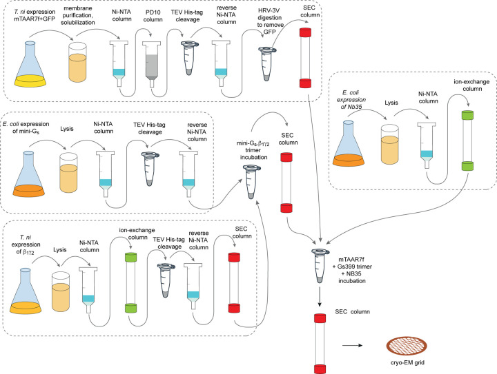
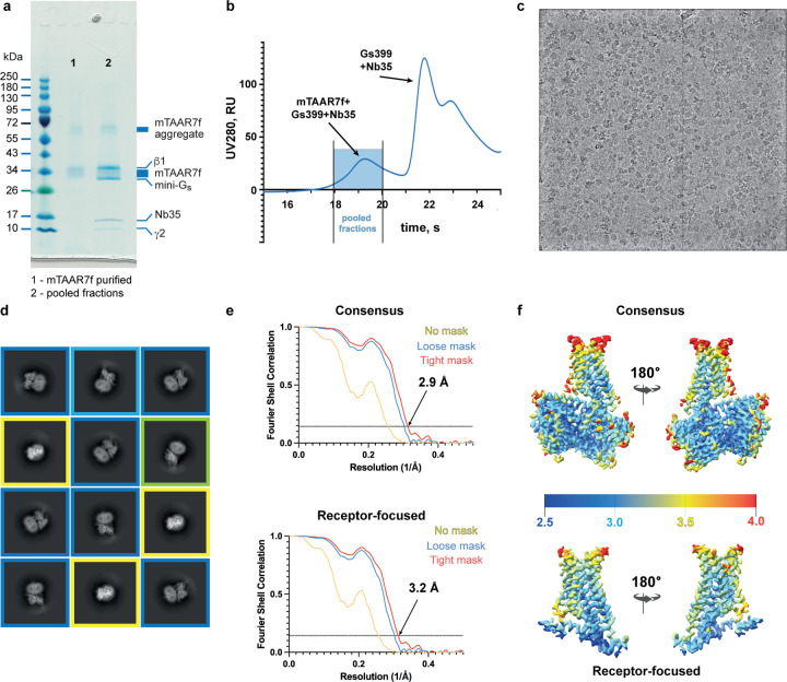
There are two main families of G protein-coupled receptors that detect odours in humans, the odorant receptors (ORs) and the trace amine-associated receptors (TAARs). Their amino acid sequences are distinct, with the TAARs being most similar to the aminergic receptors such as those activated by adrenaline, serotonin and histamine. To elucidate the structural determinants of ligand recognition by TAARs, we have determined the cryo-EM structure of a murine receptor, mTAAR7f, coupled to the heterotrimeric G protein Gs and bound to the odorant N,N-dimethylcyclohexylamine (DMCH) to an overall resolution of 2.9 Å. DMCH is bound in a hydrophobic orthosteric binding site primarily through van der Waals interactions and a strong charge-charge interaction between the tertiary amine of the ligand and an aspartic acid residue. This site is distinct and non-overlapping with the binding site for the odorant propionate in the odorant receptor OR51E2. The structure, in combination with mutagenesis data and molecular dynamics simulations suggests that the activation of the receptor follows a similar pathway to that of the β-adrenoceptors, with the significant difference that DMCH interacts directly with one of the main activation microswitch residues.


Figures

Extended Data Fig. 1 |
Extended Data Fig. 1 |. mTAAR7f purification scheme.
Purification scheme for the preparation of the mTAAR7f–miniGs–Nb35 complex for structure determination by cryo-EM.
Extended Data Fig. 2 |
Extended Data Fig. 2 |. Cryo-EM of the mTAAR7f–mini-Gs–Nb35 complex and single-particle reconstruction.
a, Coomassie Blue-stained SDS-PAGE gel of purified mTAAR7f (lane 1) and pooled fractions of the mTAAR7f–miniGs–Nb35 complex after gel filtration (lane 2). Individual components are indicated. b, Gel filtration trace of the mixture of mTAAR7f with miniGs and Nb35. c, A representative cryo-EM micrograph (defocus −2.4 μm) from the collected dataset. d, Representative 2D class averages of the mTAAR7f–miniGs–Nb35 complex determined using the initial set of particles following several rounds of 2D classification. Class averages corresponding to similar particle orientations are marked with the same coloured frames: blue, side views; green, partial side view; yellow, top views. e, FSC curves of the receptor-focused and consensus reconstructions show an overall resolution of 3.2 Å and 2.9 Å, respectively, using the gold standard FSC of 0.143. f, Local resolution estimation of the receptor-focused and consensus maps of the mTAAR7f–miniGs–Nb35 as calculated by CryoSparc.
Extended Data Fig. 3 |
Extended Data Fig. 3 |. Flow chart of cryo-EM data processing.
The dataset was collected in one session (48 h) on the LMB Krios 2 equipped with Falcon 4 detector. The movies were corrected for drift, beam-induced motion and radiation damage using CryoSparc motion correction implementation. After estimation of CTF parameters, the dataset was manually curated to exclude low quality micrographs. Particles were picked using a Gaussian blob and subjected to four rounds of 2D classification, after each round only species resembling a receptor-G protein complex were retained. Particles in the best 2D classes were subjected to two rounds of heterogenous refinement in CryoSparc versus three separately generated classes corresponding to picks without any structural features (noise classes). The output particles were subjected to one round of non-uniform refinement in CryoSparc resulting in a global resolution of 3.05 Å. To perform post-processing steps in Relion, motion correction was repeated in Relion followed by particle re-extraction and realignment. Bayesian polishing was performed in Relion, and particles were also split into AFIS groups using EPU_group_AFIS script. The final step of non-uniform refinement coupled to per-particle defocus refinement and per-particle CTF refinement, including beam-tilt, trefoil and tetrafoil corrections, was performed in CryoSparc. The final model based on 172,639 particles achieved a global resolution of 2.9 Å, while the receptor-focused map achieved a global resolution of 3.2 Å. Resolution of the models after refinements was calculated with the gold-standard FSC of 0.143 in CryoSparc (Extended Data Fig. 2).
Extended Data Fig. 4 |
Extended Data Fig. 4 |. Alignment of the amino acid sequences of mTAAR7f and β2AR.
Red bars, transmembrane regions; yellow bar, amphipathic helix 8; red residues, Ballesteros Weinstein numbering system xx.50. The alignment was performed using the program MacVector.
Extended Data Fig. 5 |
Extended Data Fig. 5 |. Sequence conservation in TAARs and the OBS.
a, Amino acid residues within 3.9 Å of ligands in the mTAAR7f structure and structures of human β2AR. b, Amino acid residues within 3.9 Å of ligands in the mTAAR7f structure aligned with the equivalent residues in both human and mouse TAARs. Sequences were aligned using Clustal Omega, and the percentage of the full-length receptor sequence identity to mTAAR7f was calculated using the web-based resource BLAST. c, Conservation in TAARs of the D-R-Y motif, transmission switches (including the P-I-F motif, marked in red) and N-P-x-x-Y motifs. Sequences were aligned using Clustal Omega,. Percentage of the full-length receptor sequence identity to mTAAR7f was calculated using web-based resource BLAST
Extended Data Fig. 6 |
Extended Data Fig. 6 |. MD simulation of DMCH association to mTAAR7f.
a, Four 2.2 μsec velocity MD simulation were performed on mTAAR7f (no G protein) in the presence of ligand outside the OBS. Three examples are shown in Extended Data Fig. 8 and one example is shown here where the ligand remained stably associated with the receptor at the end of the simulation. The ligand was observed to enter the OBS rapidly. Ligand clustering analysis identified specific residues (Asp296 and Tyr308) that associated with DMCH upon initial association with the receptor. The process is plotted visually through measuring distances between DMCH and the residues in the extracellular region (Asp2966.58 and Tyr3087.35) and in the OBS (Asp1273.32 and Trp2866.48). The motion of Trp286 is monitored through the variation in its Chi2 angle. b, Three step model for the binding of DMCH into mTAAR7f. Note that this simulation was performed on mTAAR7f in an active state and might not represent fully the trajectory in an inactive state in the absence of a G protein. However, given our understanding of the role of the G protein in closing the entrance of the OBS and decreasing its volume upon G protein coupling in the βARs, then the data here may represent an underestimate of the rate of ligand association.
Extended Data Fig. 7 |
Extended Data Fig. 7 |. MD simulations of mTAAR7f and analysis of changes in activation switches.
a, Five independent MD simulations were performed either on the mTAAR7f-mini-Gs-DMCH complex (with G protein), on mTAAR7f-DMCH (no G protein) or on mTAAR7f alone (Apo). The mean GPCR backbone RMSD and the mean volume of the OBS are plotted for each simulation and found to increase significantly in the Apo simulations compared to when G protein and ligand are bound. b, Position of the transmission elements in mTAAR7f that were analysed to assess whether the receptor was remaining in the state defined by the cryo-EM structure. These included all the canonical transmission switches in Class A GPCRs. c, For each of the above simulations distances were plotted between residues that define the state of the transmission switches. No significant differences were observed in the OBS (D127-Y316 lock), but increases in distances were observed for both mTAAR7f and β2AR in the YY motif and the sodium, binding site, consistent with a tendency towards a more inactive state. No change was observed in the NPxxY motif in mTAAR7f, but β2AR changed towards a more inactive state. Changes in Chi2 angle of Trp286 in mTAAR7f show a tendency towards a more inactive state in the Apo simulation, but this is not in evidence in β2AR. Data for simulations on β2AR were obtained from GPCRmd. The error bars represent the SD and a t-test showed either no statistical difference (ns) or a statistical difference (*, p< 0.05) between data.
Extended Data Fig. 8 |
Extended Data Fig. 8 |. MD simulation of DMCH association to mTAAR7f.
Three additional 2.2 μsec velocity MD simulation are shown of mTAAR7f (no G protein) in the presence of ligand outside the OBS. The colour scheme is identical to that in Extended Data Fig. 6a. The blue area in the traces (Step3) represents where the simulated position of DMCH is similar to that in the cryo-EM structure. In Velocity 2 the ligand is stable in Step 3, but in Velocity 3 the ligand dissociates and does not re-bind. In Velocity 4 the initially adopts a pose similar to the cryo-EM structure, but then rotates away form it, until re-adopting the cryo-EM pose 1 μsec later. Panels on the left represent the first 200–400 nsec and the panels on the right show the whole 2.2 μsec simulation.
Extended Data Fig. 9 |
Extended Data Fig. 9 |. BRET data for G protein recruitment to mTAAR7f.
a, BRET ratios measured for increasing concentrations of the agonist DMCH for the wild-type mTAAR7f (M1) and six mutants. b, Values for Emax and pEC50 determined from the data in panel a, with errors given as SEM. Three experiments were performed independently with single measurements per experiment.
Fig. 1 |
Fig. 1 |. Phylogenetic analysis of selected human GPCRs and overall structure of the mTAAR7f-Gs complex.
a, Representative human receptors of GPCR families interacting with different ligand types are compared to hTAAR9, the closest human homologue of mTAAR7f (72% amino acid residue identity). The phylogenetic analysis tool from GPCRdb.org was used. b, Cryo-EM density of the entire complex. c, cartoon of the mTAAR7f-Gs complex (ribbon representation) with bound DMCH (pale blue) and CHS (pale brown) shown as spheres. The inset shows density for DMCH (pale blue) and surrounding residues (purple) in mesh. The view is from the extracellular surface and is 90° orthogonal to the receptor cartoon viewed in the membrane plane. d, The three most populated ligand binding poses derived from MD simulations conducted either in the absence or presence of the G protein (ligand orientation from the cryo-EM structure is shown in grey). The bar graph shows differences in ligand RMSD from five distinct MD simulations either with or without G protein. The error bars represent the SD and a t-test showed no statistical difference (ns) between the mean ligand RMSDs.
Figure 2. |
Figure 2. |. The mTAAR7f orthosteric binding site and comparison to other receptors.
a, Sliced surface representation of the OBS of DMCH-bound mTAAR7f, serotonin-bound 5HT4R, adrenaline-bound β2AR and propionate-bound OR51E2; ligand atoms are depicted as spheres and the structures are shown below. b, Binding pose of DMCH and details of ligand-receptor interactions. Amino acid residues ≤ 3.9 Å from the ligand are shown with polar interactions depicted as dashed lines. c, Amino acid residues in the OBS within 3.9 Å of the ligand of mTAAR7f, aminergic receptors and an odorant receptor, OR51E2: (PDB IDs; h5HT4R, 7XT8; β1AR, 7JJO; β2AR, 4LDO; h5HT1DR, 7E32; OR51E2, 8F76). Numbers refer to the Ballesteros-Weinstein naming convention. d, The relative positions of the OBS in OR51E2 and mTAAR7f are shown after superposition of the receptors. Ligands (propionate and DMCH) are shown as spheres. e, Frequency of ligand contacts as determined during MD simulations. f, G protein recruitment was assayed using BRET arising from NanoLuc-labelled receptor and Venus-labelled mini-Gs. The mean of three independent experiments performed once are shown with error bars representing the SEM (Extended Data Fig. 9).
Figure 3. |
Figure 3. |. Interactions between mini-Gs and mTAAR7f.
a, Cartoon of the mTAAR7f-Gs complex with an inset highlighting interactions between the α5 helix of mini-Gs and mTAAR7f (distance cut-off ≤ 3.9 Å). b, Comparison of amino acid contacts (distance cut-off ≤ 3.9 Å) made by the α-subunit of Gs and mTAAR7f, h5HT4R (PDB 7XT8), hβ2AR (PDB 3SN6) and OR51E2 (PDB 8F76); blue, polar contacts; grey, van der Waals contacts.
Figure 4. |
Figure 4. |. Activation switches in mTAAR7f and β2AR.
a, Conformational changes in the functional motifs are depicted in an alignment of the inactive state of hβ2AR (yellow, carazolol-bound, PDB 2RH1), an active state of hβ2AR (orange, BI-167107-bound, PDB 3SN6) and mTAAR7 (purple). b, Increase in the TM5 bulge in β2AR upon the transition from an inactive state (left panel, yellow, PDB 2RH1) to the active state (right panel, orange, PDB 4LDO). Both structures are aligned with the active structure of mTAAR7f-Gs-DMCH (purple). Hydrogen bonds between the receptors and their corresponding ligands are shown as dashed lines. c, Alignment of amino acid residues in the bulge region of aminergic GPCR representatives with mTAAR7f, hTAAR9 and hTAAR1. One amino acid in the bulge region is absent in mTAAR7f and hTAAR9.

References

    1. Firestein S. How the olfactory system makes sense of scents. Nature 413, 211–218 (2001). - PubMed
    1. Serizawa S. et al. Negative Feedback Regulation Ensures the One Receptor-One Olfactory Neuron Rule in Mouse. Science 302, 2088–2094 (2003). - PubMed
    1. Gainetdinov R. R., Hoener M. C. & Berry M. D. Trace Amines and Their Receptors. Pharmacol. Rev. 70, 549–620 (2018). - PubMed
    1. Li Q. & Liberles S. D. Odor Sensing by Trace Amine-Associated Receptors. in Chemosensory Transduction 67–80 (Elsevier, 2016). doi: 10.1016/B978-0-12-801694-7.00004-4. - DOI
    1. Rutigliano G., Accorroni A. & Zucchi R. The Case for TAAR1 as a Modulator of Central Nervous System Function. Front. Pharmacol. 8, 987 (2018). - PMC - PubMed

Publication types