

This is a preprint.
Structural alterations in the amygdala and impaired social incentive learning in a mouse model of a genetic variant associated with neurodevelopmental disorders
- PMID: 37461714
- PMCID: PMC10350205
- DOI: 10.21203/rs.3.rs-3070199/v1
Structural alterations in the amygdala and impaired social incentive learning in a mouse model of a genetic variant associated with neurodevelopmental disorders
Abstract
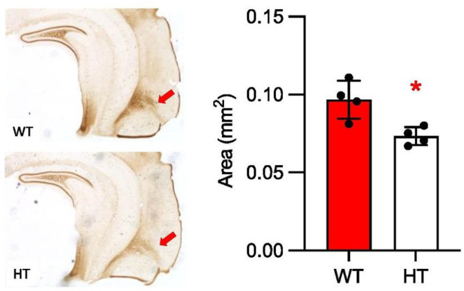
Copy number variants (CNVs) are robustly associated with psychiatric disorders and their dimensions and changes in brain structures and behavior. However, as CNVs contain many genes, the precise gene-phenotype relationship remains unclear. Although various volumetric alterations in the brains of 22q11.2 CNV carriers have been identified in humans and mouse models, it is unknown how the genes in the 22q11.2 region individually contribute to structural alterations and associated mental illnesses and their dimensions. Our previous studies have identified Tbx1, a T-box family transcription factor encoded in 22q11.2 CNV, as a driver gene for social interaction and communication, spatial and working memory, and cognitive flexibility. However, it remains unclear how TBX1 impacts the volumes of various brain regions and their functionally linked behavioral dimensions. In this study, we used volumetric magnetic resonance imaging analysis to comprehensively evaluate brain region volumes in congenic Tbx1 heterozygous mice. Our data show that the volumes of anterior and posterior portions of the amygdaloid complex and its surrounding cortical regions were reduced in Tbx1 heterozygous mice. Moreover, we examined the behavioral consequences of an altered volume of the amygdala. Tbx1 heterozygous mice were impaired for their ability to detect the incentive value of a social partner in a task that depends on the amygdala. Our findings identify the structural basis for a specific social dimension associated with loss-of-function variants of TBX1 and 22q11.2 CNV.
Keywords: 22q11.2; 22q11.2 CNV; CNV; Tbx1; amygdala; autism; schizophrenia; social incentive motivation; volumetric MRI.
Conflict of interest statement
Declaration of conflicts of interest None
Figures


Similar articles
-
Structural alterations in the amygdala and impaired social incentive learning in a mouse model of a genetic variant associated with neurodevelopmental disorders.bioRxiv [Preprint]. 2023 Aug 6:2023.06.14.545013. doi: 10.1101/2023.06.14.545013. bioRxiv. 2023. PMID: 37398198 Free PMC article. Preprint.
-
Highly demarcated structural alterations in the brain and impaired social incentive learning in Tbx1 heterozygous mice.Mol Psychiatry. 2025 May;30(5):1876-1886. doi: 10.1038/s41380-024-02797-x. Epub 2024 Oct 27. Mol Psychiatry. 2025. PMID: 39463450 Free PMC article.
-
Tbx1, a gene encoded in 22q11.2 copy number variant, is a link between alterations in fimbria myelination and cognitive speed in mice.Mol Psychiatry. 2022 Feb;27(2):929-938. doi: 10.1038/s41380-021-01318-4. Epub 2021 Nov 5. Mol Psychiatry. 2022. PMID: 34737458 Free PMC article.
-
Modeling and Predicting Developmental Trajectories of Neuropsychiatric Dimensions Associated With Copy Number Variations.Int J Neuropsychopharmacol. 2019 Aug 1;22(8):488-500. doi: 10.1093/ijnp/pyz026. Int J Neuropsychopharmacol. 2019. PMID: 31135887 Free PMC article. Review.
-
Effects of copy number variations on brain structure and risk for psychiatric illness: Large-scale studies from the ENIGMA working groups on CNVs.Hum Brain Mapp. 2022 Jan;43(1):300-328. doi: 10.1002/hbm.25354. Epub 2021 Feb 21. Hum Brain Mapp. 2022. PMID: 33615640 Free PMC article. Review.
References
Publication types
Grants and funding
LinkOut - more resources
Full Text Sources
