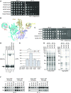
Ncs2* mediates in vivo virulence of pathogenic yeast through sulphur modification of cytoplasmic transfer RNA
- PMID: 37462076
- PMCID: PMC10450187
- DOI: 10.1093/nar/gkad564
Ncs2* mediates in vivo virulence of pathogenic yeast through sulphur modification of cytoplasmic transfer RNA
Abstract
Fungal pathogens threaten ecosystems and human health. Understanding the molecular basis of their virulence is key to develop new treatment strategies. Here, we characterize NCS2*, a point mutation identified in a clinical baker's yeast isolate. Ncs2 is essential for 2-thiolation of tRNA and the NCS2* mutation leads to increased thiolation at body temperature. NCS2* yeast exhibits enhanced fitness when grown at elevated temperatures or when exposed to oxidative stress, inhibition of nutrient signalling, and cell-wall stress. Importantly, Ncs2* alters the interaction and stability of the thiolase complex likely mediated by nucleotide binding. The absence of 2-thiolation abrogates the in vivo virulence of pathogenic baker's yeast in infected mice. Finally, hypomodification triggers changes in colony morphology and hyphae formation in the common commensal pathogen Candida albicans resulting in decreased virulence in a human cell culture model. These findings demonstrate that 2-thiolation of tRNA acts as a key mediator of fungal virulence and reveal new mechanistic insights into the function of the highly conserved tRNA-thiolase complex.
© The Author(s) 2023. Published by Oxford University Press on behalf of Nucleic Acids Research.
Figures


Similar articles
-
A highly conserved tRNA modification contributes to C. albicans filamentation and virulence.Microbiol Spectr. 2024 May 2;12(5):e0425522. doi: 10.1128/spectrum.04255-22. Epub 2024 Apr 8. Microbiol Spectr. 2024. PMID: 38587411 Free PMC article.
-
Sod1-deficient cells are impaired in formation of the modified nucleosides mcm5s2U and yW in tRNA.RNA. 2024 Nov 18;30(12):1586-1595. doi: 10.1261/rna.080181.124. RNA. 2024. PMID: 39322276 Free PMC article.
-
Transfer RNA modification and infection - Implications for pathogenicity and host responses.Biochim Biophys Acta Gene Regul Mech. 2018 Apr;1861(4):419-432. doi: 10.1016/j.bbagrm.2018.01.015. Epub 2018 Jan 31. Biochim Biophys Acta Gene Regul Mech. 2018. PMID: 29378328 Review.
-
Ubiquitin-related modifier Urm1 acts as a sulphur carrier in thiolation of eukaryotic transfer RNA.Nature. 2009 Mar 12;458(7235):228-32. doi: 10.1038/nature07643. Epub 2009 Jan 14. Nature. 2009. PMID: 19145231
-
Messenger RNA transport in the opportunistic fungal pathogen Candida albicans.Curr Genet. 2017 Dec;63(6):989-995. doi: 10.1007/s00294-017-0707-6. Epub 2017 May 16. Curr Genet. 2017. PMID: 28512683 Free PMC article. Review.
Cited by
-
Molecular basis for thiocarboxylation and release of Urm1 by its E1-activating enzyme Uba4.Nucleic Acids Res. 2024 Dec 11;52(22):13980-13995. doi: 10.1093/nar/gkae1111. Nucleic Acids Res. 2024. PMID: 39673271 Free PMC article.
-
tRNA thiolation optimizes appressorium-mediated infection by enhancing codon-specific translation in Magnaporthe oryzae.Nucleic Acids Res. 2025 Jan 7;53(1):gkae1302. doi: 10.1093/nar/gkae1302. Nucleic Acids Res. 2025. PMID: 39777460 Free PMC article.
-
tRNA hypomodification facilitates 5-fluorocytosine resistance via cross-pathway control system activation in Aspergillus fumigatus.Nucleic Acids Res. 2025 Jan 24;53(3):gkae1205. doi: 10.1093/nar/gkae1205. Nucleic Acids Res. 2025. PMID: 39711467 Free PMC article.
-
A highly conserved tRNA modification contributes to C. albicans filamentation and virulence.Microbiol Spectr. 2024 May 2;12(5):e0425522. doi: 10.1128/spectrum.04255-22. Epub 2024 Apr 8. Microbiol Spectr. 2024. PMID: 38587411 Free PMC article.
-
tRNA binding to Kti12 is crucial for wobble uridine modification by Elongator.Nucleic Acids Res. 2025 Apr 10;53(7):gkaf296. doi: 10.1093/nar/gkaf296. Nucleic Acids Res. 2025. PMID: 40226916 Free PMC article.
References
-
- Fisher M.C., Garner T.W.J. Chytrid fungi and global amphibian declines. Nat. Rev. Microbiol. 2020; 18:332–343. - PubMed
-
- Hoyt J.R., Kilpatrick A.M., Langwig K.E.. Ecology and impacts of white-nose syndrome on bats. Nat. Rev. Microbiol. 2021; 19:196–210. - PubMed
-
- Pérez J.C Fungi of the human gut microbiota: roles and significance. Int. J. Med. Microbiol. 2021; 311:151490. - PubMed
-
- Wisplinghoff H., Bischoff T., Tallent S.M., Seifert H., Wenzel R.P., Edmond M.B.. Nosocomial bloodstream infections in US hospitals: analysis of 24,179 cases from a prospective nationwide surveillance study. Clin. Infect. Dis. 2004; 39:309–317. - PubMed
-
- Sinha H., David L., Pascon R.C., Clauder-Münster S., Krishnakumar S., Nguyen M., Shi G., Dean J., Davis R.W., Oefner P.J.et al. .. Sequential elimination of major-effect contributors identifies additional quantitative trait loci conditioning high-temperature growth in yeast. Genetics. 2008; 180:1661–1670. - PMC - PubMed

