. 2023 Nov;36(6):531-541.
doi: 10.1111/pcmr.13112. Epub 2023 Jul 18.

Dynamic regulation of chromatin accessibility during melanocyte stem cell activation

Affiliations

Dynamic regulation of chromatin accessibility during melanocyte stem cell activation

Seoyeon Lee et al. Pigment Cell Melanoma Res. 2023 Nov.

Abstract

Melanocyte stem cells (McSCs) of the hair follicle are necessary for hair pigmentation and can serve as melanoma cells of origin when harboring cancer-driving mutations. McSCs can be released from quiescence, activated, and undergo differentiation into pigment-producing melanocytes during the hair cycle or due to environmental stimuli, such as ultraviolet-B (UVB) exposure. However, our current understanding of the mechanisms regulating McSC stemness, activation, and differentiation remains limited. Here, to capture the differing possible states in which murine McSCs can exist, we sorted melanocyte nuclei from quiescent (telogen) skin, skin actively producing hair shafts (anagen), and skin exposed to UVB. With these sorted nuclei, we then utilized single-nucleus assay for transposase-accessible chromatin with high-throughput sequencing (snATAC-seq) and characterized three melanocyte lineages: quiescent McSCs (qMcSCs), activated McSCs (aMcSCs), and differentiated melanocytes (dMCs) that co-exist in all three skin conditions. Furthermore, we successfully identified differentially accessible genes and enriched transcription factor binding motifs for each melanocyte lineage. Our findings reveal potential gene regulators that determine these melanocyte cell states and provide new insights into how aMcSC chromatin states are regulated differently under divergent intrinsic and extrinsic cues. We also provide a publicly available online tool with a user-friendly interface to explore this comprehensive dataset, which will provide a resource for further studies on McSC regulation upon natural or UVB-mediated stem cell activation.

Keywords: UVB; melanocyte stem cells; single-nucleus ATAC-sequencing; skin.


Figures

Figure 1:
Figure 1:
Melanocyte lineages throughout the hair cycle and after UVB irradiation. a,b, Experimental approach to enrich melanocytes in differing states at particular stages of the hair follicle cycle (telogen and anagen) and after UVB irradiation of the skin. a, Schematic of melanocyte cell states in the telogen and anagen phase of the hair follicle cycle and after UVB exposure. b, Experimental timeline for collecting telogen, anagen, and UVB-irradiated skin. c, GFP lineage tracing and DAPI staining in telogen, anagen, and after UVB exposure. Location of melanocyte lineages are shown by expression of Dct-GFP (Dct-rtTA; Tre-H2B-GFP) at the hair bulge (telogen), hair bulb (anagen), and infundibulum (UVB irradiated). Scale bar: 100μm.
Figure 2:
Figure 2:
Single-nucleus chromatin accessibility of melanocyte lineage cells. a, UMAP plot of 14,674 cells (2,585 telogen cells, 5,474 anagen cells, 6,615 UVB cells) that passed quality control. Eight different clusters were identified by unbiased clustering and colors represent the five major cell types. b, UMAP plot of Dct gene accessibility. c, Genome tracks showing aggregated snATAC-seq profiles near the Dct promoter in each cluster. d, UMAP plot of 5,695 cells from the three melanocyte clusters. Colors represent three unique melanocyte lineage cell clusters. e, Normalized gene accessibility of stemness and differentiation genes. Positive accessibility indicates that the gene is more accessible than the mean, and negative accessibility indicates that the gene is less accessible than the mean across all samples. f, Bar plots showing the proportion of melanocyte subtypes in telogen, anagen, and telogen after UVB exposure. g,h, Differential cell abundance analysis using Milo (Dann et al., 2022) of cell neighborhood changes in the anagen phase (g) or after UVB exposure (h) compared to telogen phase. The size of circles (Nhood size) represents the number of cells in a neighborhood; the thickness of lines (overlap size) indicates the number of cells shared between neighborhoods. The circles are colored by the log-fold differences (logFC) between groups. Purple-colored points indicate that more cells in that neighborhood were found in the anagen phase (g) or after UVB exposure (h). White circles indicate that the neighborhoods are not differentially abundant in one group (FDR 10%).
Figure 3:
Figure 3:
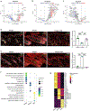
Differentially accessible genes and transcription factor motifs in three melanocyte lineages. a-c, Differentially accessible gene analysis of each melanocyte lineage cell cluster compared to other melanocyte lineage cells. Positive log2-fold change indicates increased accessibility in qMcSC (a), dMC (b), and aMcSC (c). d,e, Immunostaining of Lamb1 (d) and Ago2 (e) in telogen, anagen and after UVB exposure (left), and quantification (right). Melanocyte lineages are shown by Dct-GFP labeling in the hair bulge and hair bulb. DAPI (grey), Dct-GFP (green), and Lamb1 (red). n=4 for each group. f, Gene ontology (GO) enrichment for biological processes for the top 50 significantly more accessible genes. ****, ***, and ** indicate p-value < 0.0001, 0.001, and 0.01, respectively. Statistical test: one-way ANOVA. Scale bar: 100μm (d,e). g, Heatmap showing the enrichment of transcription factor binding motifs in each melanocyte lineage.
Figure 4:
Figure 4:
Enrichment of TFAP2A binding motifs in aMcSCs. a, Representative images of TFAP2A immunofluorescent staining on telogen, anagen, and UVB-irradiated DG dorsal skin sections (left), and quantification (right) n=4 per group. **,***,**** indicates pvalue < 0.01, 0.001,0.0001 respectively. Statistics test: one-way ANOVA. Scale bar: 100um. b, Number of TFAP2A binding sites from publicly available mouse immortalized melanocyte ChIP-seq data (Seberg et al., 2017) compared to peaks enriched in aMcSC versus qMcSC (left) and dMC (right). Gene ontology (GO) enrichment terms and FDR for biological process using the nearest genes (<100kb) to the peaks enriched in aMcSC and having TFAP2A motifs.

References

    1. Adam RC, Yang H, Ge Y, Infarinato NR, Gur-Cohen S, Miao Y, Wang P, Zhao Y, Lu CP, Kim JE, Ko JY, Paik SS, Gronostajski RM, Kim J, Krueger JG, Zheng D, Fuchs E, 2020. NFI transcription factors provide chromatin access to maintain stem cell identity while preventing unintended lineage fate choices. Nat. Cell Biol. 22, 640–650. doi:10.1038/s41556-020-0513-0 - DOI - PMC - PubMed
    1. Behrens J, von Kries JP, Kühl M, Bruhn L, Wedlich D, Grosschedl R, Birchmeier W, 1996. Functional interaction of beta-catenin with the transcription factor LEF-1. Nature 382, 638–642. doi:10.1038/382638a0 - DOI - PubMed
    1. Botchkareva NV, Botchkarev VA, Gilchrest BA, 2003. Fate of melanocytes during development of the hair follicle pigmentary unit. J. Investig. Dermatol. Symp. Proc. 8, 76–79. doi:10.1046/j.1523-1747.2003.12176.x - DOI - PubMed
    1. Brombin A, Simpson DJ, Travnickova J, Brunsdon H, Zeng Z, Lu Y, Young AIJ, Chandra T, Patton EE, 2022. Tfap2b specifies an embryonic melanocyte stem cell that retains adult multifate potential. Cell Rep. 38, 110234. doi:10.1016/j.celrep.2021.110234 - DOI - PMC - PubMed
    1. Chou WC, Takeo M, Rabbani P, Hu H, Lee W, Chung YR, Carucci J, Overbeek P, Ito M, 2013. Direct migration of follicular melanocyte stem cells to the epidermis after wounding or UVB irradiation is dependent on Mc1r signaling. Nat. Med. 19, 924–929. doi:10.1038/nm.3194 - DOI - PMC - PubMed

Publication types

Associated data

LinkOut - more resources