. 2023 Jul 18;14(1):131.
doi: 10.1007/s12672-023-00725-z.

RIPK1 is aberrantly expressed in multiple B-cell cancers and implicated in the underlying pathogenesis

Affiliations

RIPK1 is aberrantly expressed in multiple B-cell cancers and implicated in the underlying pathogenesis

Baoyu Wu et al. Discov Oncol. .

Abstract

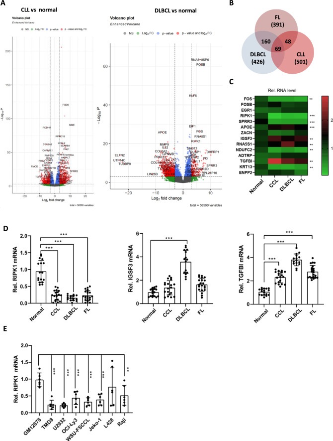
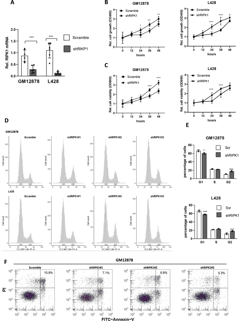
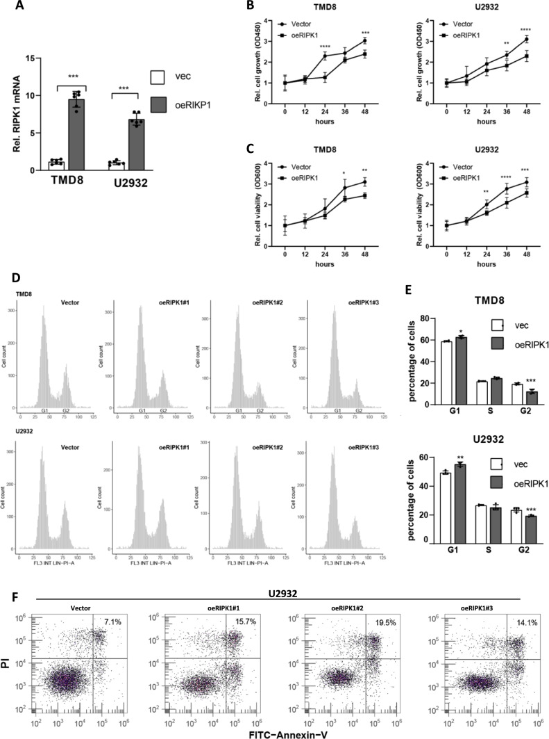
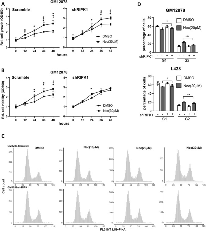
According to the latest epidemiology of the US, B-cell cancers account for > 3% of all new cancer cases and > 80% of non-Hodgkin lymphomas. However, the disease-modifying small molecular drug suitable for most B-cell cancers is still lacking. RIPK1 (receptor-interacting serine/threonine-protein kinase 1) has been observed to be dysregulated and implicated in the pathogenesis of multiple solid cancers, of which, however, the roles in blood cancers are quite unclear. In our study, to identify multi-function targets for B-cell cancer treatment, we reanalyzed a public transcriptomic dataset from the database of Gene Expression Omnibus, which includes CD19+ B-cell populations from 6 normal donors and patients of 5 CLL, 10 FL, and 8 DLBCL. After overlapping three groups (CLL vs. normal, FL vs. normal, and DLBCL vs. normal) of differentially expressed genes (DEGs), we obtained 69 common DEGs, of which 3 were validated by real-time quantitative PCR, including RIPK3, IGSF3, TGFBI. Interestingly, we found that the loss function of RIPK1 significantly increases the proliferation and viability of GM12878 cells (a normal human B lymphocyte cell line). Consistently, overexpression of RIPK1 in TMD8 and U2932 cells effectively inhibited cell proliferation and growth. More importantly, modifying RIPK1 kinase activity by a small molecule (such as necrostain-1, HOIPIN-1, etc.) alters the cell growth status of B-cell lymphoma, showing that RIPK1 exhibits anti-tumor activity in the context of B-cell lymphoma. Taken together, we consider that RIPK1 may be a potential target in the clinical application of B-cell lymphoma (including CLL, DLBCL, and FL) treatment.

Keywords: B-cell cancers; HOIPIN-1; Necrostain-1; RIPK1.


Conflict of interest statement

The authors declare no competing interests.

Figures

Fig. 1
Fig. 1
RIPK1 is down-regulated in tumor cells of lymphoma patients. A Volcano plot of DEGs of CLL and DLBCL vs. normal donors, respectively. Chronic lymphocytic leukemia (CCL, N = 5) and diffuse large B-cell patients (DLBCL, N = 8) vs. normal donors (N = 6). The cutoff was set at p-adjust < 0.001 and log2 fold change > 3. B Venn gram of three groups of DEGs. CLL and DLBCL vs. normal donor from 1A, FL vs. normal donor from S1B, all the genes in detail were listed in Table 3. C Real-time PCR estimated the mRNA level of the top 14 highly expressed DEGs in B. The CD19+ B-cells of 17 normal donors, 19 CCL, 15 DLBCL, and 21 FL were pooled together, respectively, and then were subjected to real-time PCR. The color shows the mean of the relative expression of each group. N = 3. **P < 0.01 and ***P < 0.001 by one-way ANOVA. D Real-time PCR estimated the mRNA level of 3 DEGs in CD19+ B-cells from the indicated participants. Samples are the same with C. **P < 0.01 and ***P < 0.001 by student’s t-test. E Real-time PCR estimated the mRNA level of RIPK1 in 8 lines of B-cells. Normal human B lymphocyte cell line (GM12878), B-cell non-Hodgkin lymphoma (OCI-LY3, JEKO-1, RAJI, TMD8, U2932, WSU-FSCCL), and a Hodgkin's lymphoma cell line (L428). N = 6, ***P < 0.001 by student’s t-test
Fig. 2
Fig. 2
Loss-function of RIPK1 contributes to the cell growth of lymphocytes A Real-time PCR estimated the RIPK1 knockdown efficiency in GM12878 and L428 cells. Scramble, scramble shRNA without a specific target; shRIPK1, a pool of three independent shRNAs targeting RIPK1. N = 6, ***P < 0.001 by student’s t-test. B CCK8 assay determined the cell growth of GM12878 and L428 cell lines. Cells in A were seeded onto 96-well plates, and 12 h later, cell growth status was detected every 12 h. N = 3, *P < 0.05, **P < 0.01, ***P < 0.001 by two-way ANOVA. C Cell viability of GM12878 and L428 cell lines. Cells in A were seeded onto 96-well plates, and 12 h later, cell viability was detected every 12 h. N = 3, *P < 0.05, **P < 0.01 by two-way ANOVA. DE. PI staining determined the cell cycle distribution of GM12878 and L428 cell lines. ~ 5000 cells cultured for 36 h were collected for PI staining. shRIPK1#1/2/3 indicates independent shRNA targets of RIPK1. Scr, scramble shRNA; N = 6, ***P < 0.001 by student’s t-test. E Annexin V-FITC/PI double-labeled FACS determined the cell death of GM12878 cell lines. Cells in D were used for experiments
Fig. 3
Fig. 3
Ectopic RIPK1 in lymphocytes suppresses cell growth A Real-time PCR estimated the RIPK1 knockdown efficiency in TMD8 and U2932 cells. N = 6, ***P < 0.001 by student’s t-test. B CCK8 assay determined cell proliferation of TMD8 and U2932 cell lines. Cells in A were seeded onto 96-well plates, and 12 h later, cell growth status was detected every 12 h. Vector, empty vector; oeRIPK1, RIPK1 overexpression. N = 3, *P < 0.05, **P < 0.01, ***P < 0.001 by two-way ANOVA. C Cell viability of TMD8 and U2932 cell lines. Cells in A were seeded onto 96-well plates, and 12 h later, cell growth status was detected every 12 h. Vector, empty vector; oeRIPK1, RIPK1 overexpression. N = 3, **P < 0.01, ***P < 0.001 by two-way ANOVA. DE. PI staining determined the cell cycle distribution of TMD8 and U2932 cell lines. ~ 5000 cells cultured for 36 h were collected for PI staining. oeRIPK1#1/2/3 indicates three duplications of RIPK1 overexpressed cell lines. Vector, empty vector; oeRIPK1, RIPK1 overexpression. N = 6, *P < 0.05, **P < 0.01, ***P < 0.001 by student’s t-test F. Annexin V-FITC/PI double-labeled FACS determined the cell death of U2932 cell lines. Cells in B were used for experiments.
Fig. 4
Fig. 4
RIPK1 inhibitor promotes the proliferation of lymphoma cells A CCK8 assay determined cell proliferation of GM12878 lines. Cells were seeded onto 96-well plates and pre-treated with Nec (30 μM) for 24 h, and then, cell growth status was detected by CCK8 assay every 12 h. N = 3, *P < 0.05, **P < 0.01, ***P < 0.001 by two-way ANOVA. B Cell viability of GM12878 cell lines. Cells in A were seeded onto 96-well plates and pre-treated with Nec (30 μM) for 24 h, and then, cell growth status was detected every 12 h. N = 3, *P < 0.05, **P < 0.01, ***P < 0.001 by two-way ANOVA. C-D. PI staining C determined the cell cycle distribution of GM12878 cell lines and the statistical results D. ~ 5000 cells were pre-treated with Nec (30 μM) for 24 h and then collected for PI staining. N = 6, *P < 0.05, ***P < 0.001 by student’s t-test
Fig. 5
Fig. 5
HOIPIN-1 inhibits the proliferation of lymphoma cells by blocking RIPK1 function A. CCK8 assay determined cell proliferation of TMD8 lines. Cells were seeded onto 96-well plates and pre-treated with HOIPIN-1 (HOI, 10 μM) for 24 h, and then, cell growth status was detected by CCK8 assay every 12 h. N = 3, *P < 0.05, **P < 0.01, ***P < 0.001 by two-way ANOVA. B Cell viability of TMD8 cell lines. Cells in A were seeded onto 96-well plates and pre-treated with HOI (10 μM) for 24 h, and then, cell viability status was detected every 12 h. N = 3, *P < 0.05, **P < 0.01, ***P < 0.001 by two-way ANOVA. CD. PI staining (C) determined the cell cycle distribution of TMD8 cell lines and the statistical results (D). ~ 5000 cells were pre-treated with HOI at the indicated dose for 24 h and then collected for PI staining. N = 6, *P < 0.05, ***P < 0.001 one-way ANOVA

References

    1. Armitage JO, et al. Non-HODGKIN LYMPHOMA. The Lancet. 2017;390(10091):298–310. doi: 10.1016/S0140-6736(16)32407-2. - DOI - PubMed
    1. Siegel RL, Miller KD, Jemal A. Cancer statistics, 2016. CA Cancer J Clin. 2016;66(1):7–30. doi: 10.3322/caac.21332. - DOI - PubMed
    1. Hanel W, Epperla N. Evolving therapeutic landscape in follicular lymphoma: a look at emerging and investigational therapies. J Hematol Oncol. 2021;14(1):104. doi: 10.1186/s13045-021-01113-2. - DOI - PMC - PubMed
    1. Wong DP, et al. A BAFF ligand-based CAR-T cell targeting three receptors and multiple B cell cancers. Nat Commun. 2022;13(1):217. doi: 10.1038/s41467-021-27853-w. - DOI - PMC - PubMed
    1. Ashida H, Sasakawa C, Suzuki T. A unique bacterial tactic to circumvent the cell death crosstalk induced by blockade of caspase-8. EMBO J. 2020;39(17):e104469. doi: 10.15252/embj.2020104469. - DOI - PMC - PubMed