. 2023 Jul 25;120(30):e2216658120.
doi: 10.1073/pnas.2216658120. Epub 2023 Jul 18.

MicroRNA-335-5p suppresses voltage-gated sodium channel expression and may be a target for seizure control

Affiliations

MicroRNA-335-5p suppresses voltage-gated sodium channel expression and may be a target for seizure control

Mona Heiland et al. Proc Natl Acad Sci U S A. .

Abstract

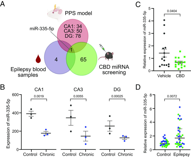
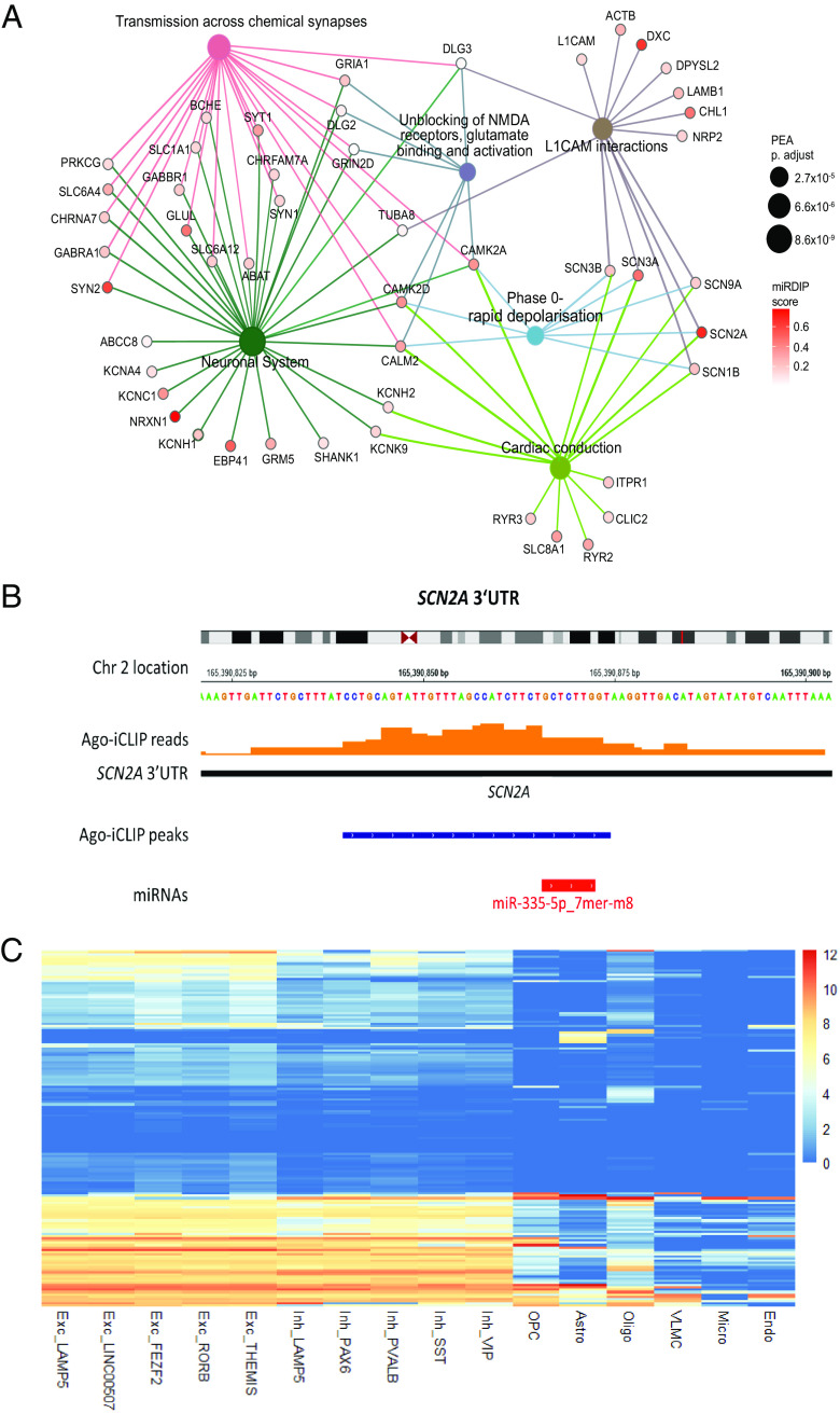
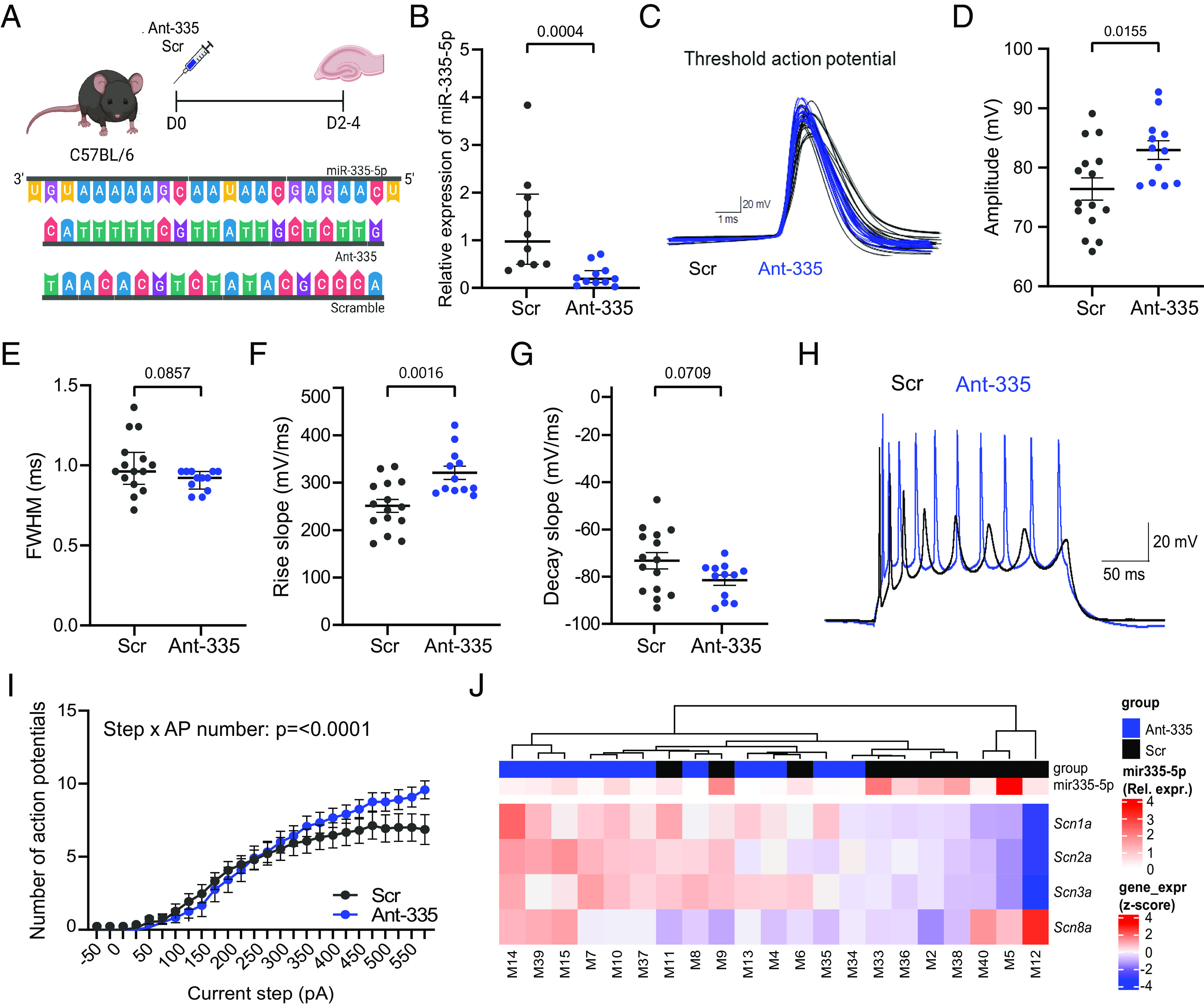
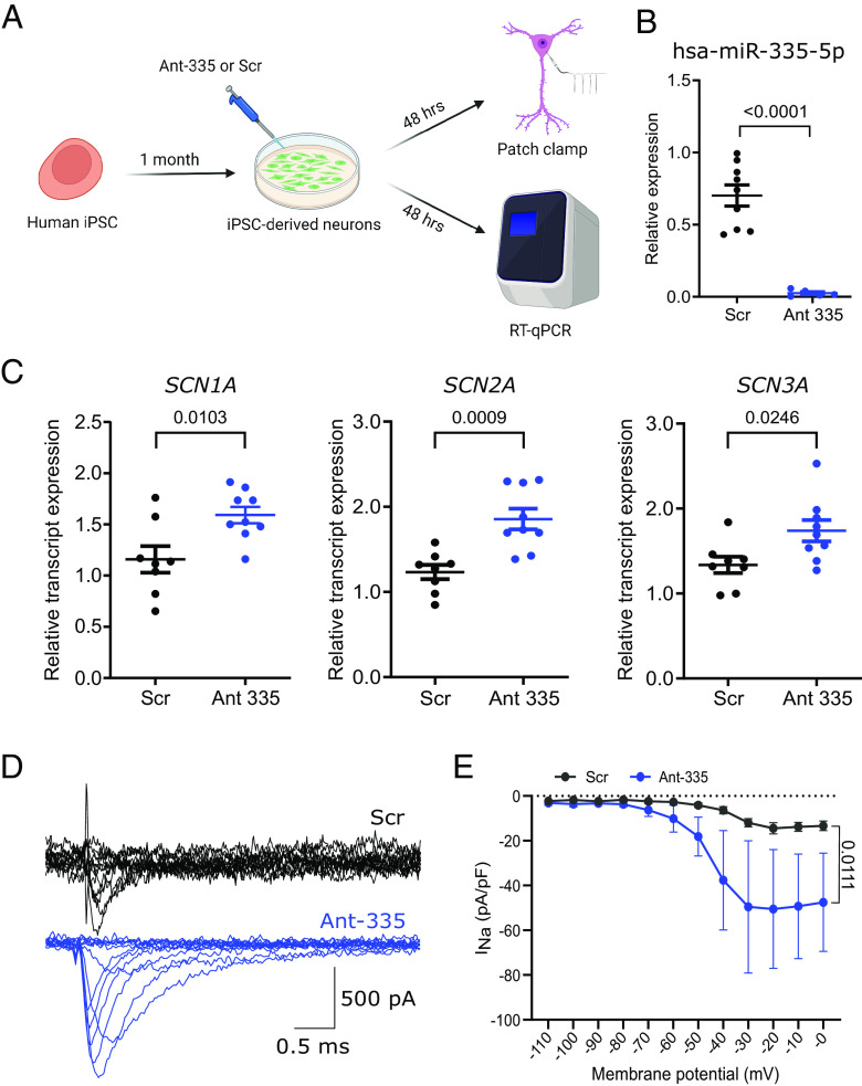
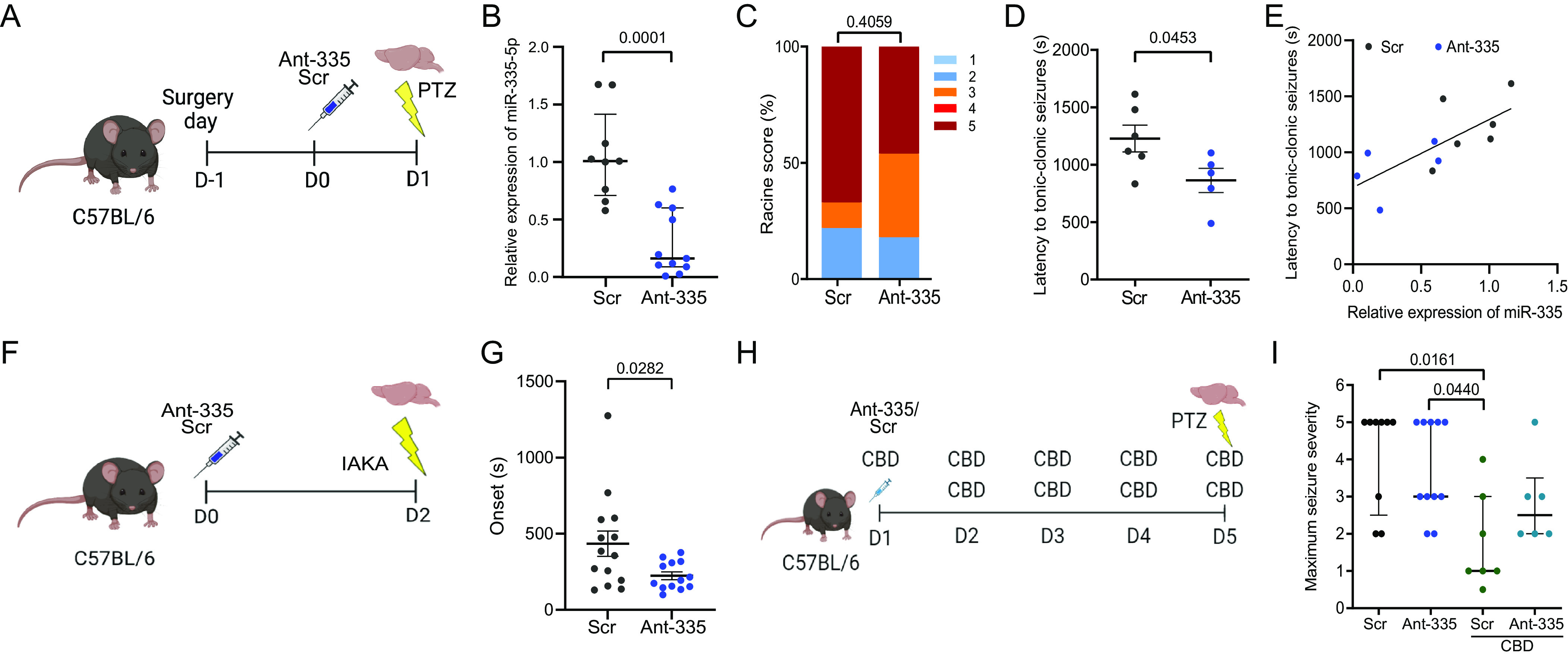
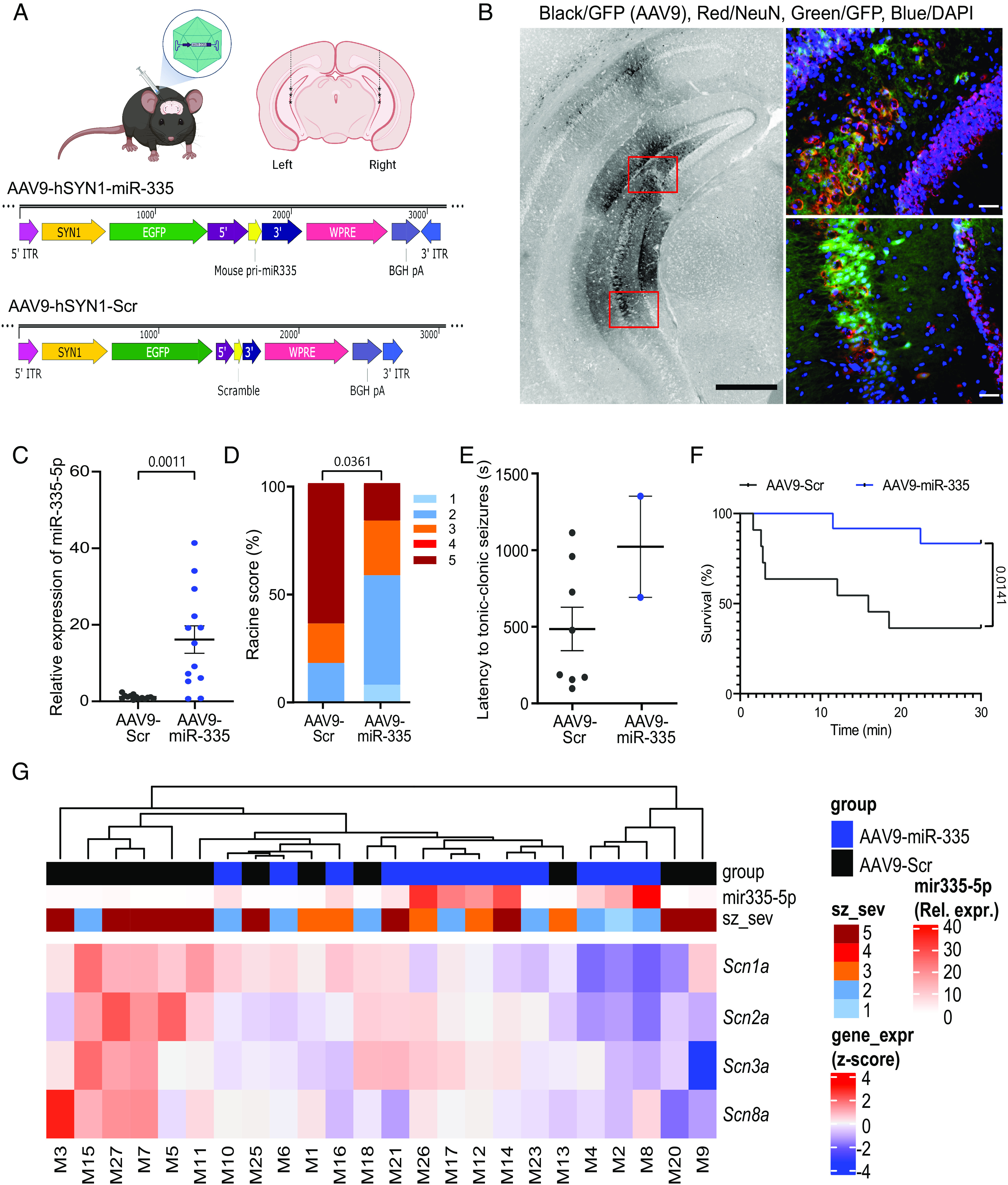
There remains an urgent need for new therapies for treatment-resistant epilepsy. Sodium channel blockers are effective for seizure control in common forms of epilepsy, but loss of sodium channel function underlies some genetic forms of epilepsy. Approaches that provide bidirectional control of sodium channel expression are needed. MicroRNAs (miRNA) are small noncoding RNAs which negatively regulate gene expression. Here we show that genome-wide miRNA screening of hippocampal tissue from a rat epilepsy model, mice treated with the antiseizure medicine cannabidiol, and plasma from patients with treatment-resistant epilepsy, converge on a single target-miR-335-5p. Pathway analysis on predicted and validated miR-335-5p targets identified multiple voltage-gated sodium channels (VGSCs). Intracerebroventricular injection of antisense oligonucleotides against miR-335-5p resulted in upregulation of Scn1a, Scn2a, and Scn3a in the mouse brain and an increased action potential rising phase and greater excitability of hippocampal pyramidal neurons in brain slice recordings, consistent with VGSCs as functional targets of miR-335-5p. Blocking miR-335-5p also increased voltage-gated sodium currents and SCN1A, SCN2A, and SCN3A expression in human induced pluripotent stem cell-derived neurons. Inhibition of miR-335-5p increased susceptibility to tonic-clonic seizures in the pentylenetetrazol seizure model, whereas adeno-associated virus 9-mediated overexpression of miR-335-5p reduced seizure severity and improved survival. These studies suggest modulation of miR-335-5p may be a means to regulate VGSCs and affect neuronal excitability and seizures. Changes to miR-335-5p may reflect compensatory mechanisms to control excitability and could provide biomarker or therapeutic strategies for different types of treatment-resistant epilepsy.

Keywords: adeno-associated virus; antisense oligonucleotides; drug resistance; epilepsy; noncoding RNA.


Conflict of interest statement

RCSI University of Medicine and Health Sciences (D.C.H., G.M., and M.H.) reports the European Patent Application No. EP21198390.3 “Modulation of microRNA-335-5p for the treatment of sodium channelopathies.”

Figures

Fig. 1.
Fig. 1.
MiR-335-5p as a common miRNA in experimental and human epilepsy, and in response to CBD. (A) MiR-335-5p emerged as the only common miRNA from interrogating tissue and biofluid miRNA datasets from a rodent model and epilepsy patients, and from CBD-treated mice. (B) Expression profile of miR-335-5p within the hippocampal circuit of the rodent epilepsy model with lower levels in the chronic epilepsy phase (n = 3/group). CA1: P = 0.0019, CA3: P = 0.0055, DG: P = 0.00025; pairwise comparison using DESeq2. (C) Relative expression of miR-335-5p after CBD treatment (n = 15/group). P = 0.0404, unpaired t test. (D) MiR-335-5p plasma levels in treatment-resistant epilepsy patients and control from two different clinical centers (n = 26 to 27/group). Green = samples from hospital in Dublin; blue = samples from hospital in Marburg, see ref. (26). P = 0.0072, Mann–Whitney U test. CA = cornu ammonis region, CBD = cannabidiol, DG = dentate gyrus, PPS = perforant path stimulation. Graphics in (A) were produced using Biorender.
Fig. 2.
Fig. 2.
Targets of miR-335-5p are predominately expressed in neurons and play an important role in pathways of neuronal excitability. (A) Top six significantly enriched Reactome pathways (sized according to p-value) from pathway enrichment analysis of the mRNA targets of miR-335-5p (miR-335-5p targets from target identification pipeline). The miR-335-5p mRNA targets associated with each pathway are colored according to miRDiP integrated score (Materials and Methods). Links between pathways and mRNA nodes are colored according to the pathway. (B) iCLIP data from human epilepsy patients identified that the SCN2A gene, which is located in chromosome 2, encodes a transcript with a target site for miR-335-5p. The orange bars represent the aggregated iCLIP reads across all the patient samples that map along the SCN2A gene. (C) Heatmap of the expression levels of miR-335-5p iCLIP targets across different cell types in the human primary motor cortex. Values are shown as trimmed means of Log2(CPM+1)(CPM = Counts Per Million). (Trimmed mean = average expression of the middle 50% of the data for each gene and cell type). Exc = Excitatory Neurons, Inh = Inhibitory Neurons, OPC = Oligodendrocyte Precursor Cells, Astro = Astrocytes, Oligo = Oligodendrocytes, VLMC = Vascular Leptomeningeal Cells, Micro = Microglia, Endo = Endothelial Cells. Excitatory and inhibitory neurons are further divided in subtypes according to the expression of the specified marker genes. Data were sourced from the Allen Brain Atlas (https://portal.brain-map.org/).
Fig. 3.
Fig. 3.
Effect of miR-335-5p inhibition on hippocampal biophysics and expression of voltage-gated sodium channel in naïve mice. (A) Schematic shows the experimental design. Briefly, adult mice were injected i.c.v. with either Ant-335 or a scrambled (Scr) sequence (sequences shown below schematic) and brain slices were prepared after 2 to 4 d for ex vivo electrophysiology. (B) Relative expression of miR-335-5p measured 48 h after i.c.v. injection of Ant-335 or scramble. P = 0.0004, Mann–Whitney U test. (C) Overlay of the threshold action potentials obtained from Ant-335- (blue) and scramble- (black) treated hippocampal CA1 pyramidal neurons. (D) Ant-335 increased action potential amplitude. P = 0.0155, unpaired t test. (E) Action potential full-width half maximum (FWHM) was not changed after Ant-335 treatment. P = 0.0857, Mann–Whitney U test. (F) The rising phase was characterized by a larger rise slope in Ant-335 treated neurons. P = 0.0016, unpaired t test. (G) Decay slope was not altered. P = 0.0709, unpaired t test. (H) Representative raw traces from Ant-335- (blue) and scramble- (black) treated neurons. (I) Ant-335 increased the maximum firing frequency of pyramidal neurons, which were able to fire above the plateau seen in control neurons, P <= 0.0001, two-way repeated measures ANOVA) Electrophysiology: n = 12 to 15 neurons per group. (J) Heatmap showing relative mRNA expression of the various VGSCs in the hippocampus after i.c.v. injection of Ant-335 or Scr (n = 10 to 11/group, mRNA expression levels (gene_expr) are z-score normalized). Expression levels of miR-335-5p [from (B)] of the corresponding animals are also shown to observe links between expression levels of miR-335-5p and expression of VGSCs. Correction for multiple comparisons was applied on electrophysiological data and α was adjusted to 0.01. Graphics in (A) were produced using Biorender.
Fig. 4.
Fig. 4.
Effect of miR-335-5p inhibition on voltage-gated sodium currents in iPSC-derived neurons. (A) Schematic shows the experimental design. Human iPSC-derived neurons were differentiated for 4 wk and treated for 48 h with Ant-335 or scramble (Scr). Voltage-gated sodium currents were then recorded in voltage-clamp mode from transfected neurons. (B) qRT-PCR performed on separate iPSC-derived neurons confirmed robust inhibition of miR-335-5p and (C) corresponding de-repression of VGSC transcripts. MiR-335-5p: P = <0.0001, unpaired t test; SCN1A: P = 0.0103, unpaired t test; SCN2A: P = 0.0009, unpaired t test; SCN3A: P = 0.0246, unpaired t test. N = 8 to 9/group. (D) Representative traces of voltage-gated sodium current elicited by step depolarizations ranging from −100 mV to 0 mV in 10 mV increments for 100 ms from a holding potential of −110 mV. (E) Current density curve showing the peak current density at different holding potentials (n = 6 to 8 neurons). P = 0.0111, two-way repeated measures ANOVA. Graphics in (A) were produced using Biorender.
Fig. 5.
Fig. 5.
miR-335-5p inhibition increases seizure susceptibility in mouse models. (A) Schematic shows the experimental design for PTZ testing. Mice were equipped with a guide cannula and injected i.c.v. with either Ant-335 or a scrambled sequence. Seizures were induced 24 h after the treatment via an i.p. injection of PTZ (80 mg/kg). (B) Relative expression of miR-335-5p measured 24 h after i.c.v. injection of Ant-335 or scramble (n = 9 to 11/group). P = 0.0001, Mann–Whitney U test. (C) Racine scale scores assessed seizure severity in the two groups (n = 9 to 11/group). P = 0.4059, Fisher’s exact test. (D) Latency to tonic-clonic seizures was decreased in animals treated with Ant-335 which reached Racine scale 5. P = 0.0453, unpaired t test. (E) Positive correlation between miR-335-5p expression and latency to tonic-clonic seizures. r = 0.7230, R2 = 0.5228, P = 0.0119. (F) Experimental design for intra-amygdala kainic acid (IAKA) testing. (G) Ant-335 reduced the time to onset of status epilepticus (SE) following IAKA (n = 13 to 14/group). P = 0.0282, unpaired t test. (H) Experimental design to test the impact of Ant-335 on seizure susceptibility in CBD-treated mice. (I) Ant-335 counteracts the reduction in seizure severity observed with CBD treatment (n = 7 to 11/group). The Kruskal–Wallis test with Dunn’s multiple comparisons test. Graphics in (A, F, and H) were produced using Biorender.
Fig. 6.
Fig. 6.
Effects of miR-335-5p overexpression on PTZ-induced seizures. (A) Schematic shows the experimental design. Briefly, adult C57 mice were injected intra-hippocampally with either an AAV9 containing the pri-miRNA sequence of miR-335-5p or a scrambled sequence. Seizures were induced 2 wk after the treatment via an i.p. injection of PTZ (75 mg/kg). (B) Expression of AAV9 (black) in the ventral part of the hippocampus. (Scale bar magnification: 500 µm.) Sections marked in red are shown in the small panels besides showing the expression of AAV9 in CA3 pyramidal neurons. NeuN = red, GFP (AAV9) = green, DAPI = blue. (Scale bar magnifications: 50 µm.) (C) Relative expression of miR-335-5p in the ventral hippocampus after intra-hippocampal injection of AAV9-miR-335 or control AAV9 (n = 9/group). P = 0.0011, unpaired t test. (D) Racine’s scale scores assessed seizure severity in the two groups (n = 11 to 12/group). P = 0.0361, Fisher’s exact test. (E) Latency to tonic-clonic seizures was not affected by the overexpression of miR-335-5p. (F) Kaplan–Meier survival analysis for the two different treatment groups. AAV9-Scr group showed a survival rate of 36% whereas animals overexpressing miR-335-5p had a survival rate of 83.3%. P = 0.0141, Mantel-Cox test. (G) Heatmap showing relative mRNA expression of the various VGSCs in the ventral hippocampus after intra-hippocampal injection of AAV9-miR-335 or control AAV9 [n = 12 per group, mRNA expression levels (gene_expr) are z-score normalized]. Seizure severity scores (sz_sev) and relative expression levels of miR-335-5p are also shown. Levels of miR-335-5p (from C) and seizure severity (from D) of the corresponding animals were incorporated to observe links between expression levels of miR-335-5p, seizure severity and expression of VGSCs. Graphics in (A) were produced using Biorender.

Comment in

Similar articles

Cited by

References

    1. Fisher R. S., et al. , ILAE official report: A practical clinical definition of epilepsy. Epilepsia 55, 475–482 (2014). - PubMed
    1. Löscher W., Klein P., The Pharmacology and Clinical Efficacy of Antiseizure Medications: From Bromide Salts to Cenobamate and Beyond. CNS Drugs 35, 935–963 (2021). - PMC - PubMed
    1. Brodie M. J., Sodium Channel Blockers in the Treatment of Epilepsy. CNS Drugs 31, 527–534 (2017). - PubMed
    1. Meisler M. H., Hill S. F., Yu W., Sodium channelopathies in neurodevelopmental disorders. Nat. Rev. Neurosci. 22, 152–166 (2021). - PMC - PubMed
    1. Steel D., Symonds J. D., Zuberi S. M., Brunklaus A., Dravet syndrome and its mimics: Beyond SCN1A. Epilepsia 58, 1807–1816 (2017). - PubMed

Publication types