Review
. 2023 Jul;29(7):332-345.
doi: 10.1089/ten.tec.2023.0082.

Three-Dimensional Printing Bioceramic Scaffolds Using Direct-Ink-Writing for Craniomaxillofacial Bone Regeneration

Affiliations
Review

Three-Dimensional Printing Bioceramic Scaffolds Using Direct-Ink-Writing for Craniomaxillofacial Bone Regeneration

Vasudev Vivekanand Nayak et al. Tissue Eng Part C Methods. 2023 Jul.

Abstract

Defects characterized as large osseous voids in bone, in certain circumstances, are difficult to treat, requiring extensive treatments which lead to an increased financial burden, pain, and prolonged hospital stays. Grafts exist to aid in bone tissue regeneration (BTR), among which ceramic-based grafts have become increasingly popular due to their biocompatibility and resorbability. BTR using bioceramic materials such as β-tricalcium phosphate has seen tremendous progress and has been extensively used in the fabrication of biomimetic scaffolds through the three-dimensional printing (3DP) workflow. 3DP has hence revolutionized BTR by offering unparalleled potential for the creation of complex, patient, and anatomic location-specific structures. More importantly, it has enabled the production of biomimetic scaffolds with porous structures that mimic the natural extracellular matrix while allowing for cell growth-a critical factor in determining the overall success of the BTR modality. While the concept of 3DP bioceramic bone tissue scaffolds for human applications is nascent, numerous studies have highlighted its potential in restoring both form and function of critically sized defects in a wide variety of translational models. In this review, we summarize these recent advancements and present a review of the engineering principles and methodologies that are vital for using 3DP technology for craniomaxillofacial reconstructive applications. Moreover, we highlight future advances in the field of dynamic 3D printed constructs via shape-memory effect, and comment on pharmacological manipulation and bioactive molecules required to treat a wider range of boney defects.

Keywords: 3D printing; bone regeneration; in vivo; preclinical; regenerative medicine; scaffold.


Conflict of interest statement

No competing financial interests exist.

Figures

FIG. 1.
FIG. 1.
(a) Biomimetic scaffold design CAD workflow schematic detailing layer-by-layer scaffold slicing and rendering (image not to scale), (b) schematic of location of unilateral calvarial defect (dashed red circle highlighting the region of interest), (c) inferior view of 3D diagram of printed calvarial scaffold, and (d) intraoperative image of fit-and-fill reconstruction of calvarial defect with scaffold. CAD, computer-aided design.
FIG. 2.
FIG. 2.
(a) Schematic of robocasting machine (3D Inks LLC, Tulsa, OK) used to assemble bioceramic scaffolds, (b) magnified set-up of extrusion nozzles (3 × ) capable of codepositing multiple materials, (c) schematic of various scaffold macrogeometries capable of being extruded through DIW 3D printing of shear thinning bioceramic colloidal gels (images not to scale). DIW, direct inkjet writing.
FIG. 3.
FIG. 3.
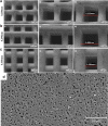
Scanning electron microscope images of 3D printed β-TCP scaffolds printed with different nozzle diameters. (a) 0.200 mm, (b) 0.250 mm, (c) 0.330 mm. (i, ii, iii) Show capacity of DIW to fabricate scaffolds with varying pore sizes, and (d) microgeometry and surface texture of β-TCP scaffold sintered to 1100°C for 4 h., β-TCP, β-tricalcium phosphate.
FIG. 4.
FIG. 4.
(a) Three-dimensional reconstructions of scaffold (purple) in the calvarium (image not to scale), (b) degradation kinetics analysis of scaffold over 18 months used to calculate annual degradation rate of β-TCP in the calvarium, and (c) elastic modulus (E) of calvarial scaffold-regenerated bone shows no difference compared to that of native control. Error bars are 95% confidence intervals.
FIG. 5.
FIG. 5.
Nondecalcified histologic sections of (a) calvarial defect and (b) alveolar defect filled with 3DP scaffolds. Red arrows indicate biomimetic ceramic scaffolds and white arrows indicate organized bone formation within the engineered pores of the scaffolds., White dashed lines denote the ends/margins of the 3D printed scaffold. 3DP, three-dimensional printing.
FIG. 6.
FIG. 6.
(a) Surgical segment compared to scaffold (b) surgical placement of scaffold, and (c) sagittal histologic slice of scaffold in continuity with rabbit mandible. White dashed lines denote the ends/margins of the 3D printed scaffold. I, incisor; IAN, inferior alveolar nerve; T, tooth.
FIG. 7.
FIG. 7.
Histological cross section of (a) negative control defect in the radius, with the white arrows indicating the screw locations for the surgical hardware; radius defect along the long axis of the 3D printed bioceramic scaffold at the defect site at (b) 8 weeks, (c) 12 weeks, and (d) 24 weeks. The marrow (M) space is visible with yellow marrow observed at 12- and 24-week time points, and (e) nanoindentation values of hydrated bone samples at 8, 12, and 24 weeks with native radius for comparison of hardness (H) of bone (The letters represent statistically homogeneous results). H at 8 and 12 weeks had similar means, whereas the values at 24 weeks were statistically >2 previous time points and achieved statistical homogeneity values relative to native bone.
FIG. 8.
FIG. 8.
(a) Stacked plot of repetitive shape-memory cycles indicating successive shape transformations through five different shape-memory cycles. Induced strains alter the shape of the construct—the shape is “frozen in” if it is maintained constant while temperature is decreased below Tg (B–C), and “recovered” if it is once again heated above Tg (D–A), and (b) illustration of the 1-W SME process. SME, shape-memory effect. Image generated on Biorender.com
FIG. 9.
FIG. 9.
(a) SEM micrograph of scaffold augmented with rhBMP-2 with signs of suture fusion (red arrows), and (b) nondecalcified histology of calvarial scaffold shows blood vessels stained with Stevenel's blue and patent sutures with evidence of suture patency (yellow splines) adjacent to sites of reconstruction. rhBMP-2, recombinant human bone morphogenetic protein-2.

References

    1. Lanza RP. Principles of Tissue Engineering, 5th ed: Academic Press: London; 2020.
    1. Seper L, Piffkó J, Joos U, et al. . Treatment of fractures of the atrophic mandible in the elderly. J Am Geriatr Soc 2004;52(9):1583–1584. - PubMed
    1. Tovar N, Witek L, Atria P, et al. . Form and functional repair of long bone using 3D printed bioactive scaffolds. J Tissue Eng Regen Med 2018;12(9):1986–1999. - PMC - PubMed
    1. Bernardo MP, da Silva BCR, Hamouda AEI, et al. . PLA/hydroxyapatite scaffolds exhibit in vitro immunological inertness and promote robust osteogenic differentiation of human mesenchymal stem cells without osteogenic stimuli. Sci Rep 2022;12(1):2333. - PMC - PubMed
    1. Kenley RA, Yim K, Abrams J, et al. . Biotechnology and bone graft substitutes. Pharm Res 1993;10(10):1393–1401. - PubMed

Publication types

LinkOut - more resources