Larger hypothalamic volume in narcolepsy type 1
- PMID: 37463428
- PMCID: PMC10636249
- DOI: 10.1093/sleep/zsad173
Larger hypothalamic volume in narcolepsy type 1
Abstract
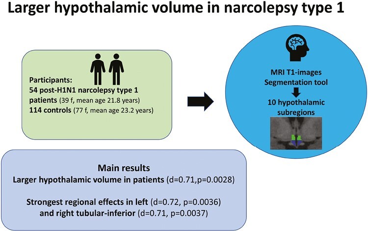
Study objectives: Narcolepsy type 1 (NT1) is a neurological sleep disorder. Postmortem studies have shown 75%-90% loss of the 50 000-70 000 hypocretin-producing neurons and 64%-94% increase in the 64 000-120 000 histaminergic neurons and conflicting indications of gliosis in the hypothalamus of NT1 patients. The aim of this study was to compare MRI-based volumes of the hypothalamus in patients with NT1 and controls in vivo.
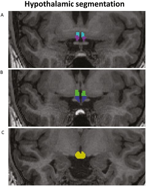
Methods: We used a segmentation tool based on deep learning included in Freesurfer and computed the volume of the whole hypothalamus, left/right part of the hypothalamus, and 10 hypothalamic subregions. We included 54 patients with post-H1N1 NT1 (39 females, mean age 21.8 ± 11.0 years) and 114 controls (77 females, mean age 23.2 ± 9.0 years). Group differences were tested with general linear models using permutation testing in Permutation Analysis of Linear Models and evaluated after 10 000 permutations, yielding two-tailed P-values. Furthermore, a stepwise Bonferroni correction was performed after dividing hypothalamus into smaller regions.
Results: The analysis revealed larger volume for patients compared to controls for the whole hypothalamus (Cohen's d = 0.71, p = 0.0028) and for the left (d = 0.70, p = 0.0037) and right part of the hypothalamus (d = 0.65, p = 0.0075) and left (d = 0.72, p = 0.0036) and right tubular-inferior (d = 0.71, p = 0.0037) hypothalamic subregions.
Conclusions: In conclusion, patients with post-H1N1 NT1 showed significantly larger hypothalamic volume than controls, in particular in the tubular-inferior subregions which could reflect several processes as previous studies have indicated neuroinflammation, gliosis, and changes in the numbers of different cell types.
Keywords: MRI; T1; hypothalamus; narcolepsy.
© The Author(s) 2023. Published by Oxford University Press on behalf of Sleep Research Society.
Figures