Review
. 2023 Jul 17;133(14):e170498.
doi: 10.1172/JCI170498.

The lung mesenchyme in development, regeneration, and fibrosis

Affiliations
Review

The lung mesenchyme in development, regeneration, and fibrosis

Elie El Agha et al. J Clin Invest. .

Abstract

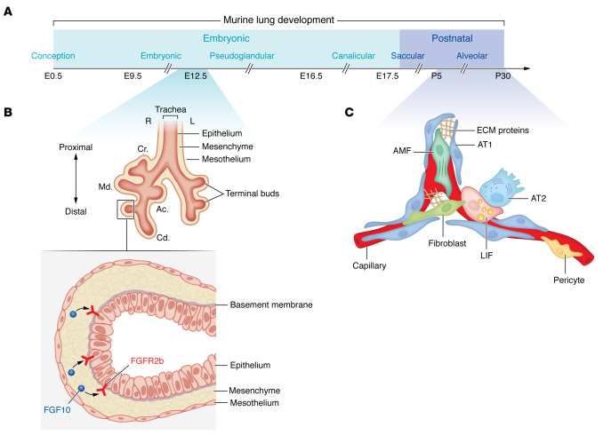
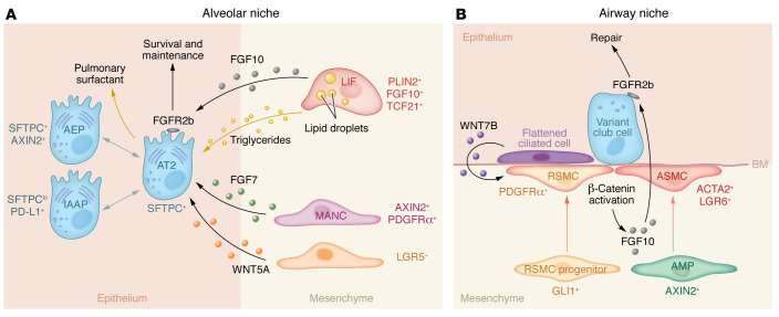
Mesenchymal cells are uniquely located at the interface between the epithelial lining and the stroma, allowing them to act as a signaling hub among diverse cellular compartments of the lung. During embryonic and postnatal lung development, mesenchyme-derived signals instruct epithelial budding, branching morphogenesis, and subsequent structural and functional maturation. Later during adult life, the mesenchyme plays divergent roles wherein its balanced activation promotes epithelial repair after injury while its aberrant activation can lead to pathological remodeling and fibrosis that are associated with multiple chronic pulmonary diseases, including bronchopulmonary dysplasia, idiopathic pulmonary fibrosis, and chronic obstructive pulmonary disease. In this Review, we discuss the involvement of the lung mesenchyme in various morphogenic, neomorphogenic, and dysmorphogenic aspects of lung biology and health, with special emphasis on lung fibroblast subsets and smooth muscle cells, intercellular communication, and intrinsic mesenchymal mechanisms that drive such physiological and pathophysiological events throughout development, homeostasis, injury repair, regeneration, and aging.


Conflict of interest statement

Conflict of interest: The authors have declared that no conflict of interest exists.

Figures

Figure 1
Figure 1. Overview of pre- and postnatal lung development.
(A) Lung development consists of an embryonic and postnatal phase. The different stages of mouse lung development are shown through P30. (B) A schematic representation of an embryonic lung at E12.5 and a corresponding sagittal section of a distal epithelial bud with the surrounding mesenchymal tissue. FGF10/FGFR2b signaling is highlighted. (C) A schematic representation of an alveolus with the constituent cell types during the alveolar stage of lung development. Cr., cranial lobe; Md., medial lobe; Cd., caudal lobe; Ac., accessory lobe; AMF, alveolar myofibroblast; AT1, alveolar epithelial type 1; AT2, alveolar epithelial type 2; LIF, lipofibroblast.
Figure 2
Figure 2. Mesenchymal-epithelial interactions in the alveolar and airway niches.
(A) AT2 and their so-far identified subclusters are shown. Among the identified niche cells are LIFs, MANCs, and LGR5+ cells. Cellular markers are also shown. (B) A WNT-FGF feedback loop mediates epithelial-mesenchymal communication during airway regeneration. AEP, alveolar epithelial progenitor; AMP, AXIN2+ myofibrogenic progenitor; ASMC, airway smooth muscle cell; AT2, alveolar epithelial type 2; BM, basement membrane; Epi, epithelium; IAAP, injury-activated alveolar progenitor; LGR5, leucine-rich repeat containing G protein–coupled receptor 5; LIF, lipofibroblast; MANC, mesenchymal alveolar niche cell; Mes, mesenchyme; RSMC, repair-supportive mesenchymal cell.

References

    1. Tsikis ST, et al. Targeting the lung endothelial niche to promote angiogenesis and regeneration: a review of applications. Front Mol Biosci. 2022;9:109336. doi: 10.3389/fmolb.2022.1093369. - DOI - PMC - PubMed
    1. Millar FR, et al. The pulmonary endothelium in acute respiratory distress syndrome: insights and therapeutic opportunities. Thorax. 2016;71(5):462–473. doi: 10.1136/thoraxjnl-2015-207461. - DOI - PubMed
    1. Cao Z, et al. Targeting of the pulmonary capillary vascular niche promotes lung alveolar repair and ameliorates fibrosis. Nat Med. 2016;22(2):154–162. doi: 10.1038/nm.4035. - DOI - PMC - PubMed
    1. Ding BS, et al. Endothelial-derived angiocrine signals induce and sustain regenerative lung alveolarization. Cell. 2011;147(3):539–553. doi: 10.1016/j.cell.2011.10.003. - DOI - PMC - PubMed
    1. Niethamer TK, et al. Defining the role of pulmonary endothelial cell heterogeneity in the response to acute lung injury. Elife. 2020;9:e53072. doi: 10.7554/eLife.53072. - DOI - PMC - PubMed

Publication types