. 2023 Sep:65:102818.
doi: 10.1016/j.redox.2023.102818. Epub 2023 Jul 13.

Dissecting the role of the NADPH oxidase NOX4 in TGF-beta signaling in hepatocellular carcinoma

Affiliations

Dissecting the role of the NADPH oxidase NOX4 in TGF-beta signaling in hepatocellular carcinoma

Rut Espinosa-Sotelo et al. Redox Biol. 2023 Sep.

Abstract

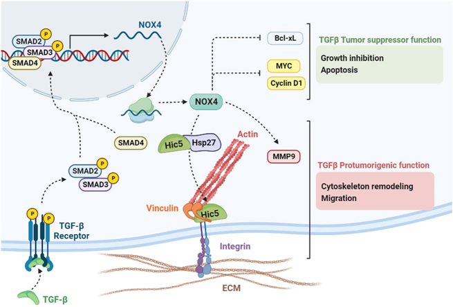
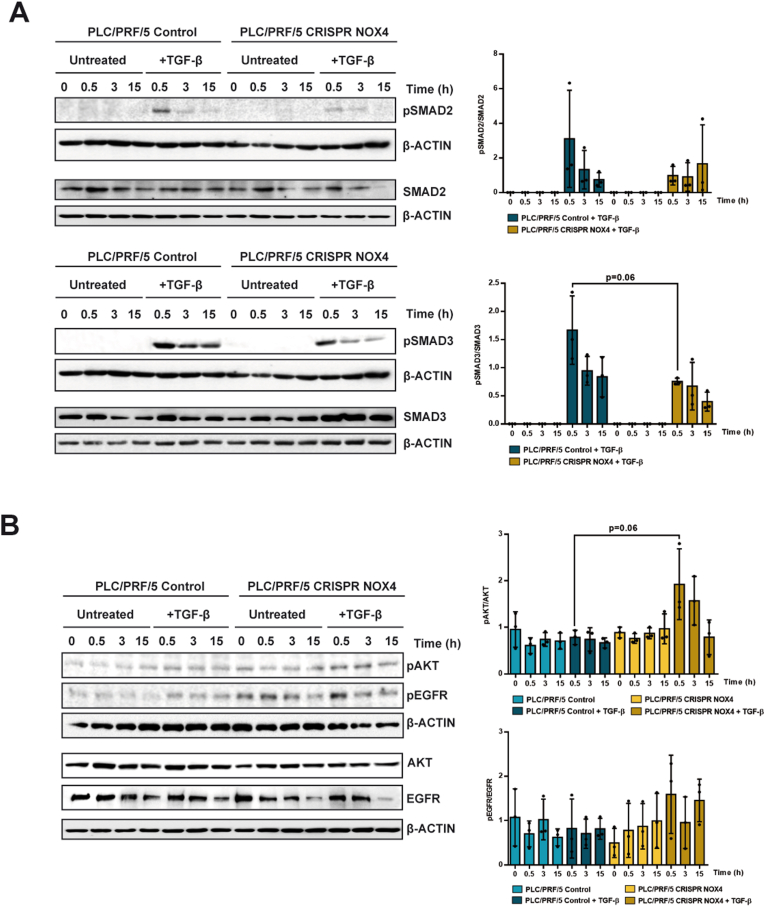
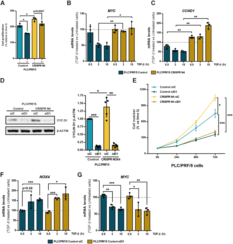
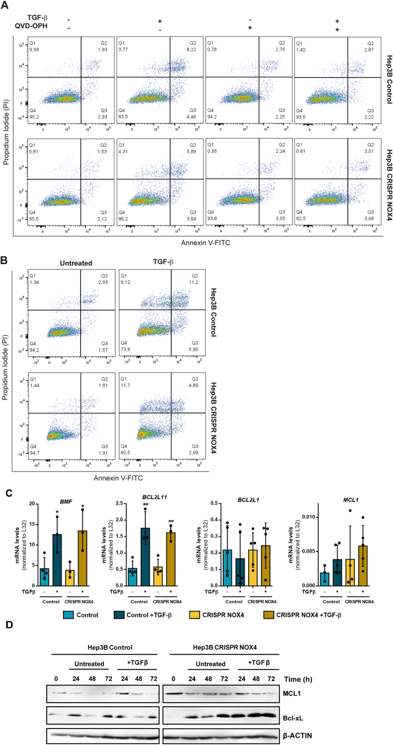
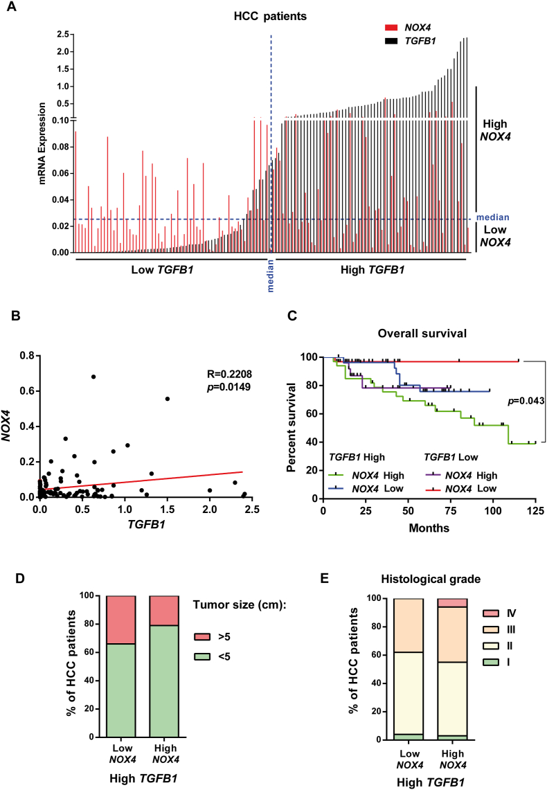
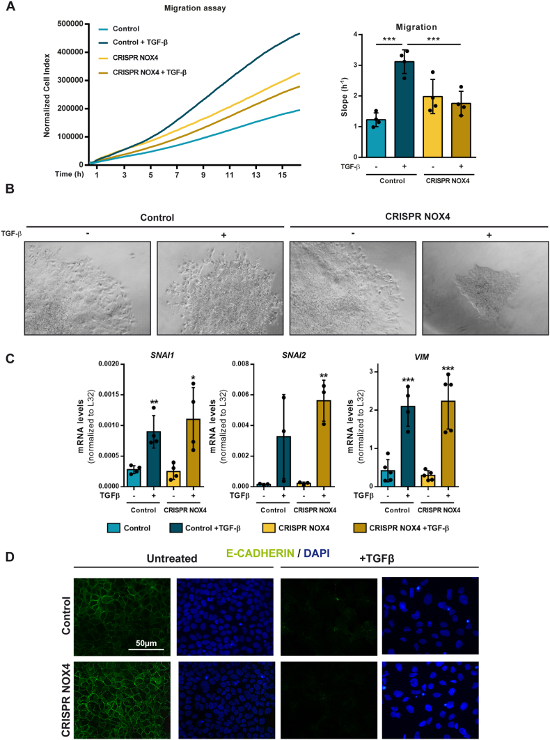
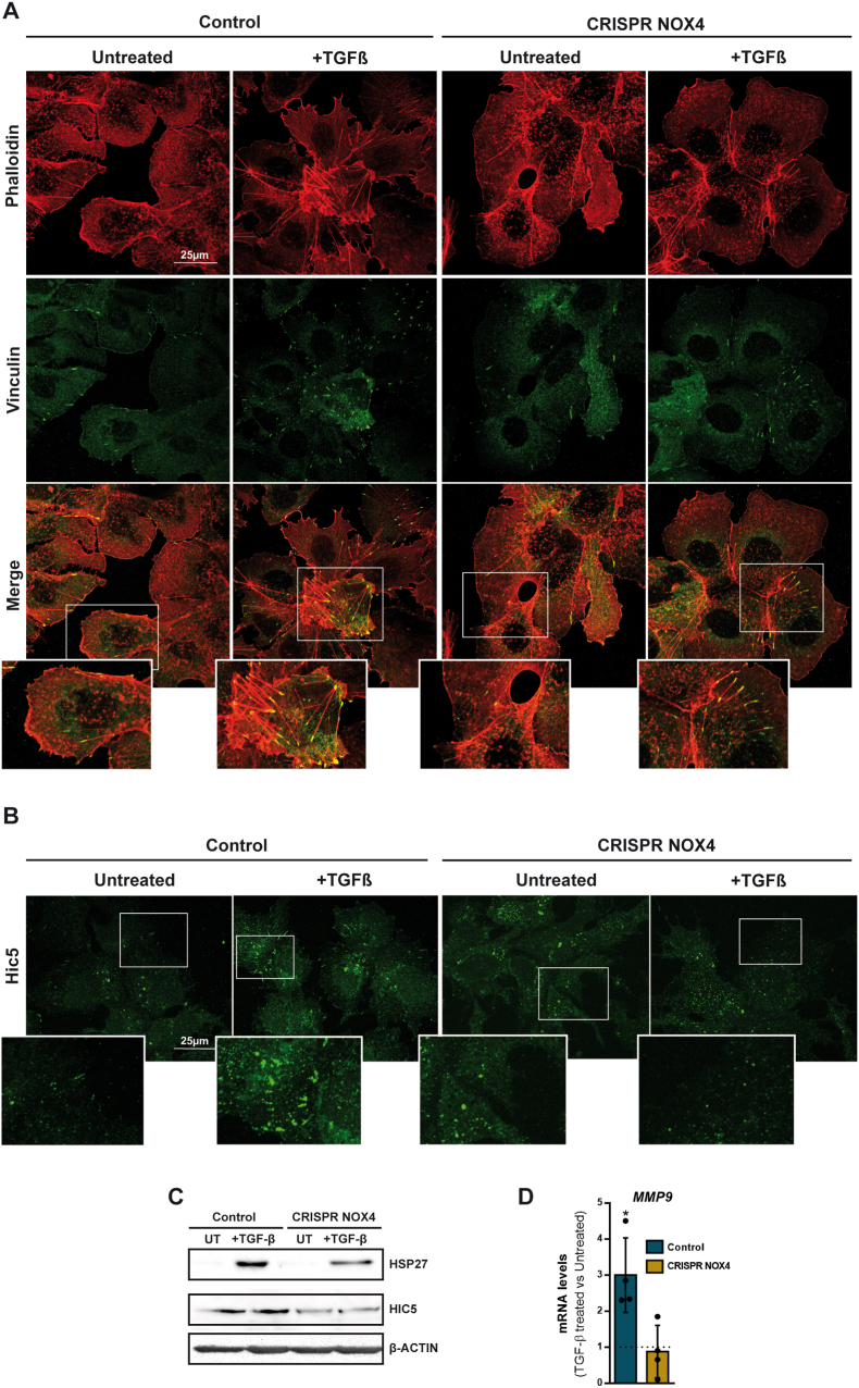
The NADPH oxidase NOX4 has been proposed as necessary for the apoptosis induced by the Transforming Growth Factor-beta (TGF-β) in hepatocytes and hepatocellular carcinoma (HCC) cells. However, whether NOX4 is required for TGF-β-induced canonical (SMADs) or non-canonical signals is not fully understood yet, neither its potential involvement in other parallel actions induced by TGF-β. In this work we have used CRISPR Cas9 technology to stable attenuate NOX4 expression in HCC cells. Results have indicated that NOX4 is required for an efficient SMAD2/3 phosphorylation in response to TGF-β, whereas non-canonical signals, such as the phosphorylation of the Epidermal Growth Receptor or AKT, are higher in NOX4 silenced cells. TGF-β-mediated inhibition of cell proliferation and viability is attenuated in NOX4 silenced cells, correlating with decreased response in terms of apoptosis, and maintenance of high expression of MYC and CYCLIN D1. These results would indicate that NOX4 is required for all the tumor suppressor actions of TGF-β in HCC. However, analysis in human HCC tumors has revealed a worse prognosis for patients showing high expression of TGF-β1-related genes concomitant with high expression of NOX4. Deepening into other tumorigenic actions of TGF-β that may contribute to tumor progression, we found that NOX4 is also required for TGF-β-induced migratory effects. The Epithelial-Mesenchymal transition (EMT) program does not appear to be affected by attenuation of NOX4 levels. However, TGF-β-mediated regulation of cytoskeleton dynamics and focal adhesions require NOX4, which is necessary for TGF-β-induced increase in the chaperone Hsp27 and correct subcellular localization of Hic-5 within focal adhesions, as well for upregulation of the metalloprotease MMP9. All these results together point to NOX4 as a key element in the whole TGF-β signaling in HCC cells, revealing an unknown role for NOX4 as tumor promoter in HCC patients presenting activation of the TGF-β pathway.

Keywords: HCC; Hepatocellular carcinoma; Liver cancer; NADPH oxidase; NOX4; TGF-Beta.


Conflict of interest statement

Declaration of competing interest The authors declare the following financial interests/personal relationships which may be considered as potential competing interests: Isabel Fabregat reports financial support was provided by Agencia Estatal de Investigación, Ministry of Science and Innovation, Spain. Rut Espinosa-Sotelo reports financial support was provided by Agencia Estatal de Investigación, Ministry of Science and Innovation, Spain. Isabel Fabregat reports financial support was provided by Asociación Española contra el Cáncer (AECC), Spain. Isabel Fabregat reports financial support was provided by Agència de Gestió d’Ajuts Universitaris i de Recerca (AGAUR, Generalitat de Catalunya). Isabel Fabregat reports financial support was provided by CIBER, National Biomedical Research Institute, Instituto de Salud Carlos III, Spain.

Figures

Image 1
Graphical abstract
Fig. 1
Fig. 1
Role of NOX4 on TGF-β-mediated canonical and non-canonical signals. Analysis made in PLC/PRF/5 (Control and CRISPR NOX4) cells, untreated or TGF-β-treated. A) Phospho-SMAD2/3 and total SMAD2/3; B) Phospho-AKT, phospho-EGFR and their corresponding total protein levels. In both cases, Western blot, β-Actin was used as loading control. Left: Representative experiments. Right: quantification of the Phospho/Total ratio in each case, after densitometric analysis of the levels taking into account the specific loading control [(Phospho)/(phospho loading)]/[(Total)/(total loading)]. Data are Mean ± SD (n = 3).
Fig. 2
Fig. 2
Role of NOX4 in the response to TGF-β in terms of cell proliferation. Analysis made in PLC/PRF/5 (Control and CRISPR NOX4) cells. A) Cell viability assay analyzed by crystal violet staining after 72h of TGF-β treatment, normalized to initial time (time 0h). Data represents mean ± SD of triplicates from one representative experiment. B) Relative MYC and C)CCND1 mRNA expression analyzed by RT-qPCR, normalized to housekeeping gene L32, after TGF-β treatment at 0, 0.5, 3 and 15 h. Represented as percentage of TGF-β treated versus untreated cells in each time. D) Cyclin D1 protein levels analyzed by Western blot after transiently transfecting either with control or Cyclin D1 specific siRNA sequences. β-Actin was used as loading control. Representative experiment (left) and densitometric analysis of Cyclin D1 levels relative to β-Actin (right). E) Cell proliferation assay analyzed by crystal violet staining after transiently transfecting with either control or Cyclin D1 specific siRNA sequences, normalized to initial time (time 0h). F) Relative NOX4 and G)MYC mRNA expression analyzed by RT-qPCR, normalized to housekeeping gene L32, in Control CRISPR PLC/PRF/5 cells transiently transfecting with either control or Cyclin D1 specific siRNA sequences: effect of TGF-β treatment at 0, 0.5, 3 and 15 h. Data are represented as percentage of TGF-β treated versus untreated cells in each time. In all the panels, data are Mean ± SD (n = 3). *p<0.05 **p<0.01 ***p<0.001. (siC: Control siRNA; siD1: Cyclin D1 siRNA). (For interpretation of the references to colour in this figure legend, the reader is referred to the Web version of this article.)
Fig. 3
Fig. 3
Role of NOX4 on TGF-β-induced apoptosis. Analysis made in Hep3B (Control and CRISPR NOX4) cells. A-B) Representative flow cytometry plots using Annexin V-FITC/PI staining for analysis of apoptosis in Hep3B (Control and CRISPR NOX4) treated with TGF-β for 48h (A) or 72h (B). In A, co-treatment with Q-VD-OPH, supporting the role of caspases in the process. C) Proapoptotic (BMF and BCL2L11) and antiapoptotic (BCL2L1 and MCL1) mRNA expression analyzed by RT-qPCR, normalized to housekeeping gene L32, after TGF-β treatment at 48 h. D) MCL1 and Bcl-xL protein levels analyzed by western blot after TGF-β treatment at 24, 48 and 72 h. β-Actin was used as loading control. In A and C data are Mean ± SD (n = 3–6). *p<0.05 **p<0.01, ***p<0.001. In B and D, a representative experiment is shown.
Fig. 4
Fig. 4
Impact of the expression of NOX4 and TGFB1 on different parameters in HCC patients. HCC patients n = 124 from HUB. A) Distribution of the patients according to the expression of TGFB1 and NOX4. B) Pearson correlation analysis between NOX4 and TGFB1 gene expression in 120 patients for which we had data of survival: TGFB1 High/NOX4 High (green: 33 patients); TGFB1 High/NOX4 Low (blue: 27 patients); TGFB1 Low/NOX4 High (purple, 28 patients); TGFB1 High/NOX4 High (red, 32 patients). C) Kaplan-Meier curve for overall survival percentage when TGFB1 and NOX4 expression are high/low. D) Percentage of HCC patients that have a tumor size bigger or smaller than 5 cm when TGFB1 expression is high, depending on their NOX4 expression. E) Percentage of HCC patients in each histological grade (I-IV) when TGFB1 expression is high, depending on their NOX4 expression. (For interpretation of the references to colour in this figure legend, the reader is referred to the Web version of this article.)
Fig. 5
Fig. 5
Role of NOX4 on TGF-β-induced cell migration and Epithelial-Mesenchymal Transition. Analysis made in Hep3B (Control and CRISPR NOX4) cells. A) Cell migration after TGF-β treatment, real-time monitored using xCELLigence system. Results expressed as Normalized Cell Index (left) or slope (h-1) (right) of the first 16 h. B) Representative images of spheroids from cells either untreated or treated with TGF-β, embedded in a collagen I matrix for 96h. C)SNAI1, SNAI2 and VIM mRNA expression analyzed by RT-qPCR, normalized to housekeeping gene L32, after 48h TGF-β treatment. D) Immunofluorescence of E-Cadherin (green) and DAPI (blue) for nuclei staining after 48h TGF-β treatment. In A-Right and C data are Mean ± SD (n = 3–4). *p<0.05 **p<0.01, ***p<0.001. In B and D, representative images are shown. Scale bar, 50 μm. (For interpretation of the references to colour in this figure legend, the reader is referred to the Web version of this article.)
Fig. 6
Fig. 6
Role of NOX4 on TGF-β-induced cytoskeleton remodeling. Analysis made in Hep3B (Control and CRISPR NOX4) cells. A) Immunofluorescence of Phalloidin (red) and Vinculin (green) after 48h TGF-β treatment. B) Immunofluorescence of Hic-5 (green) after 48h TGF-β treatment. C) Hsp27 and Hic-5 protein levels analyzed by western blot after 48 h of TGF-β treatment. β-Actin was used as loading control D)MMP9 mRNA expression analyzed by RT-qPCR, normalized to housekeeping gene L32, after 48h TGF-β treatment and represented as fold induction (TGF-β-treated versus untreated cells). In A and B, representative images are shown. Scale bar, 25 μm (A). Scale bar, 25 μm (B). In D, data are Mean ± SD (n = 4–6). *p<0.05. (For interpretation of the references to colour in this figure legend, the reader is referred to the Web version of this article.)

References

    1. Gough N.R., Xiang X., Mishra L. TGF-Β signaling in liver, pancreas, and gastrointestinal diseases and cancer. Gastroenterology. 1 de agosto de. 2021;161(2):434–452.e15. - PMC - PubMed
    1. Herranz-Itúrbide M., Peñuelas-Haro I., Espinosa-Sotelo R., Bertran E., Fabregat I. The TGF-β/NADPH oxidases axis in the regulation of liver cell biology in health and disease. Cells. 1 de septiembre de. 2021;10(9) - PMC - PubMed
    1. Carmona-Cuenca I., Roncero C., Sancho P., Caja L., Fausto N., Fernández M., et al. Upregulation of the NADPH oxidase NOX4 by TGF-beta in hepatocytes is required for its pro-apoptotic activity. Journal of Hepatology. diciembre de. 2008;49(6):965–976. - PubMed
    1. Yu J.H., Zhu B.M., Riedlinger G., Kang K., Hennighausen L. The liver-specific tumor suppressor STAT5 controls expression of the reactive oxygen species-generating enzyme NOX4 and the proapoptotic proteins PUMA and BIM in mice. Hepatology. diciembre de. 2012;56(6):2375–2386. - PMC - PubMed
    1. Caja L., Sancho P., Bertran E., Iglesias-Serret D., Gil J., Fabregat I. Overactivation of the MEK/ERK pathway in liver tumor cells confers resistance to TGF-β-induced cell death through impairing up-regulation of the NADPH oxidase NOX4. Cancer Research. 1 de octubre de. 2009;69(19):7595–7602. - PubMed

Publication types

MeSH terms