Eosinophilic esophagitis
- PMID: 37463709
- PMCID: PMC10370381
- DOI: 10.15537/smj.2023.44.7.20220812
Eosinophilic esophagitis
Abstract
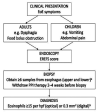
Eosinophilic esophagitis (EoE) is an atopic disease in which eosinophils infiltrate the esophageal mucosa and may result in a variety of upper gastrointestinal symptoms. Chief among these are dysphagia, heartburn, and food bolus obstruction in adults whereas children often present with abdominal pain or vomiting. Eosinophilic esophagitis is a chronic condition that if not detected and left untreated could lead to the development of subepithelial fibrosis and esophageal stenosis. The diagnosis of EoE is confirmed in a patient presenting with characteristic EoE symptoms, classic signs on endoscopy, and biopsy results showing >15 eosinophils/hpf. A number of useful treatments against EoE are currently available with new therapeutics on the horizon. The former include PPIs, topical steroids, and elimination diet; the latter comprise novel biologics including the monoclonal antibody dupilumab. All these treatments can improve symptoms and reduce esophageal eosinophil count. This brief introductory review describes the detection, diagnosis, and management of EoE.
Keywords: dupilumab; eosinophilic esophagitis; gastroesophageal reflux disease; proton pump inhibitor.
Copyright: © Saudi Medical Journal.
Figures


Similar articles
-
Endoscopic findings in patients with Schatzki rings: evidence for an association with eosinophilic esophagitis.World J Gastroenterol. 2012 Dec 21;18(47):6960-6. doi: 10.3748/wjg.v18.i47.6960. World J Gastroenterol. 2012. PMID: 23322994 Free PMC article.
-
[Diagnosis and management of eosinophilic esophagitis].Rev Med Chil. 2020 Jun;148(6):831-841. doi: 10.4067/S0034-98872020000600831. Rev Med Chil. 2020. PMID: 33480383 Spanish.
-
Eosinophilic Esophagitis: A Review.JAMA. 2021 Oct 5;326(13):1310-1318. doi: 10.1001/jama.2021.14920. JAMA. 2021. PMID: 34609446 Free PMC article. Review.
-
Eosinophilic esophagitis: Pathophysiology, diagnosis, and management.Arch Pediatr. 2019 Apr;26(3):182-190. doi: 10.1016/j.arcped.2019.02.005. Epub 2019 Mar 1. Arch Pediatr. 2019. PMID: 30827775 Review.
-
Prevalence and endoscopic features of eosinophilic esophagitis in patients with esophageal or upper gastrointestinal symptoms.J Dig Dis. 2012 Jun;13(6):296-303. doi: 10.1111/j.1751-2980.2012.00589.x. J Dig Dis. 2012. PMID: 22624552
Cited by
-
Adherence to clinical guidelines for the evaluation and management of eosinophilic esophagitis among gastroenterologists in the Arab countries.Front Pediatr. 2025 Apr 10;13:1521266. doi: 10.3389/fped.2025.1521266. eCollection 2025. Front Pediatr. 2025. PMID: 40276102 Free PMC article.
References
-
- Landres RT, Kuster GG, Strum WB.. Eosinophilic esophagitis in a patient with vigorous achalasia. J Gastroenterol 1978; 74: 1298–1301. - PubMed
Publication types
MeSH terms
Supplementary concepts
LinkOut - more resources
Full Text Sources
Medical

