A retrospective review of Mohs micrographic surgery trends over more than 10 years in Saudi Arabia
- PMID: 37463713
- PMCID: PMC10370377
- DOI: 10.15537/smj.2023.44.7.20220892
A retrospective review of Mohs micrographic surgery trends over more than 10 years in Saudi Arabia
Abstract
Objectives: To review Mohs micrographic surgery (MMS) trends in Saudi Arabia.Mohs micrographic surgery is a precise surgical technique that has been proven to have the highest cure rate with maximum normal tissue preservation. It is the treatment of choice for non-melanoma skin cancer (NMSC), especially the aggressive histopathological forms, and tumors located in high-risk regions or where tissue preservation is a mandate.
Methods: A multicentric retrospective study was performed on patients who underwent MMS between January 2010 and September 2022. The information was extracted from the database of King Saud University Medical City and Prince Sultan Military Medical City in Saudi Arabia.
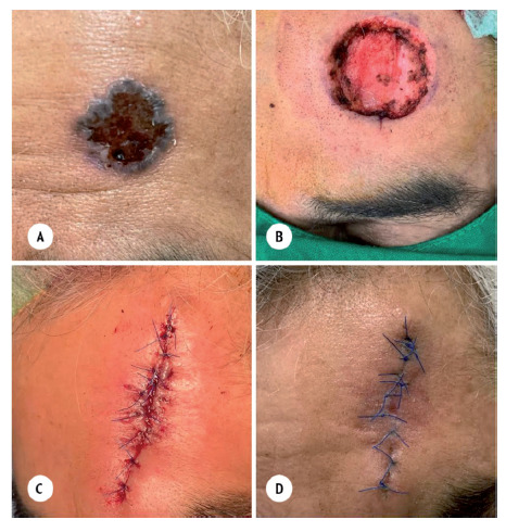
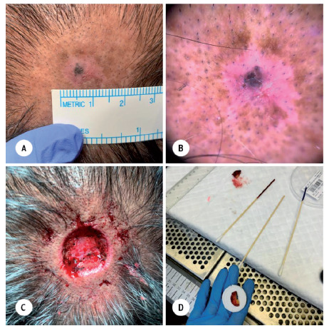
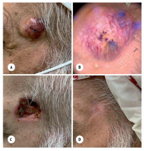
Results: A total of 70 participants were enrolled in this study. Two-thirds (67%) of the tumors that were treated using MMS were basal-cell carcinomas (BCC), 18.6% were squamous cell carcinomas (SCC), 5.7% were sebaceous carcinoma, 4.3% were dermatofibrosarcoma protuberans (DFSP), and 1.4% were rare tumors such as primary mucinous carcinoma. The most common type of reconstruction used to repair post-MMS defect was primary closure in more than half of the patients followed by secondary intention healing (20%). There were no side effects apart from a hematoma in one patient and wound infection in two patients.
Conclusion: Although MMS is still generally underutilized in Saudi Arabia, its use has increased in the last decade.
Keywords: BCC; DFSP; KSA; MMS; Mohs; Riyadh; SCC; Saudi Arabia; basosquamous carcinoma; micrographic; mucinous carcinoma; retrospective; review; sebaceous carcinoma; skin cancer; surgery.
Copyright: © Saudi Medical Journal.
Figures
Similar articles
-
Dermatofibrosarcoma protuberans: a report on 29 patients treated by Mohs micrographic surgery with long-term follow-up and review of the literature.Cancer. 2004 Jul 1;101(1):28-38. doi: 10.1002/cncr.20316. Cancer. 2004. PMID: 15221986
-
The efficacy of Mohs micrographic surgery over the traditional wide local excision surgery in the cure of dermatofibrosarcoma protuberans.Pan Afr Med J. 2019 Aug 13;33:297. doi: 10.11604/pamj.2019.33.297.17692. eCollection 2019. Pan Afr Med J. 2019. PMID: 31692830 Free PMC article. Review.
-
Wide local excision, Mohs micrographic surgery, and reconstructive options for treatment of dermatofibrosarcoma protuberans of the breast: A retrospective case series from Mayo Clinic.World J Surg Oncol. 2023 May 6;21(1):141. doi: 10.1186/s12957-023-03022-9. World J Surg Oncol. 2023. PMID: 37147611 Free PMC article.
-
Dermatofibrosarcoma protuberans: wide local excision vs. Mohs micrographic surgery.Cancer Treat Rev. 2008 Dec;34(8):728-36. doi: 10.1016/j.ctrv.2008.06.002. Epub 2008 Aug 5. Cancer Treat Rev. 2008. PMID: 18684568 Review.
-
A comparison between Mohs micrographic surgery and wide surgical excision for the treatment of dermatofibrosarcoma protuberans.J Am Acad Dermatol. 1996 Jul;35(1):82-7. J Am Acad Dermatol. 1996. PMID: 8682970 Review.
Cited by
-
Factors Affecting Dental Implant Failure: A Retrospective Analysis.Healthcare (Basel). 2025 Jun 6;13(12):1356. doi: 10.3390/healthcare13121356. Healthcare (Basel). 2025. PMID: 40565383 Free PMC article.
References
-
- Bolognia JL, Schaffer JV, Cerroni L.. Dermatology: 2-Volume Set. 4th ed. Elsevier Health Sciences; 2017.
-
- Prickett KA, Ramsey ML. Mohs Micrographic Surgery. Stat Pearls Publishing; 2023. - PubMed
Publication types
MeSH terms
LinkOut - more resources
Full Text Sources
Medical
Research Materials