. 2023 Jul 18;14(1):3900.
doi: 10.1038/s41467-023-39615-x.

Conformational restriction shapes the inhibition of a multidrug efflux adaptor protein

Affiliations

Conformational restriction shapes the inhibition of a multidrug efflux adaptor protein

Benjamin Russell Lewis et al. Nat Commun. .

Abstract

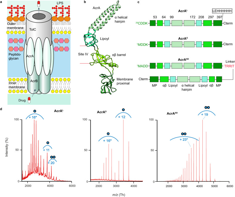
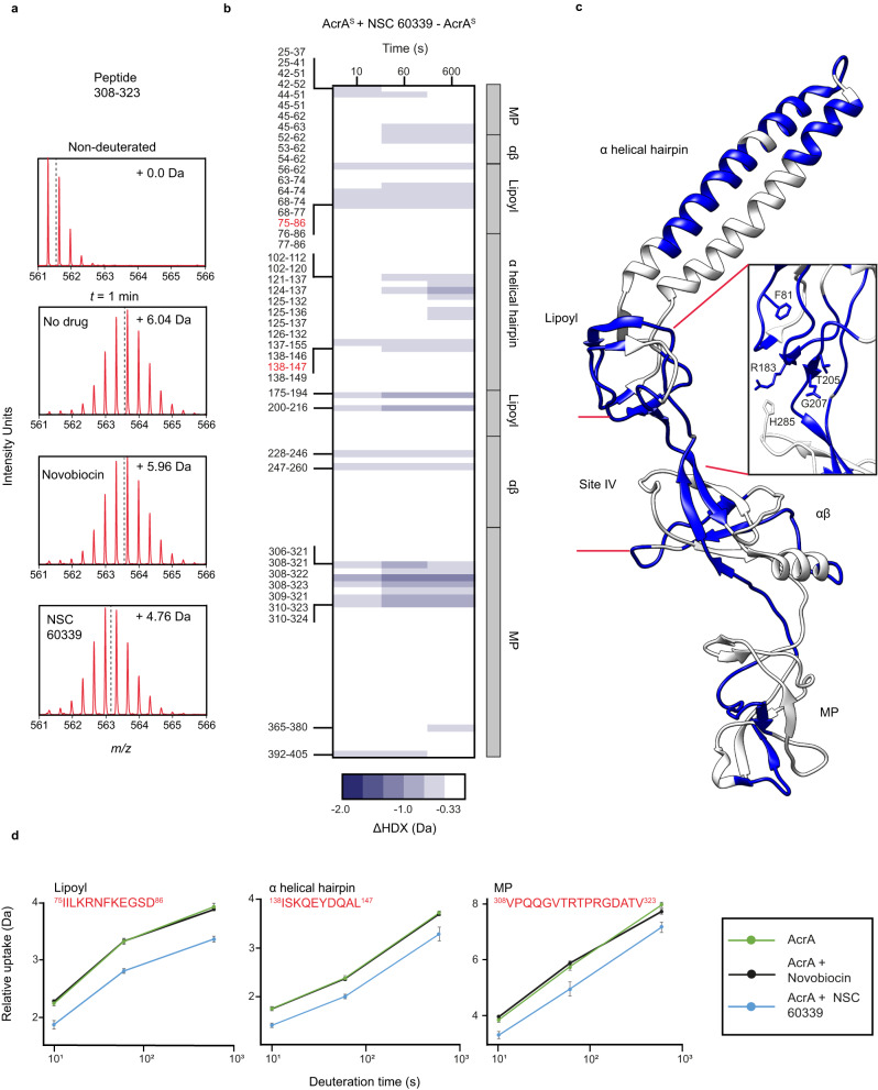
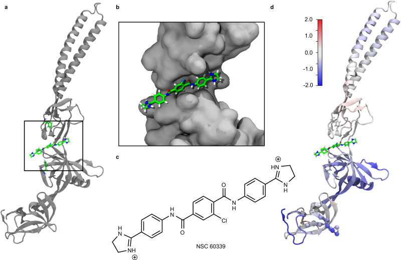
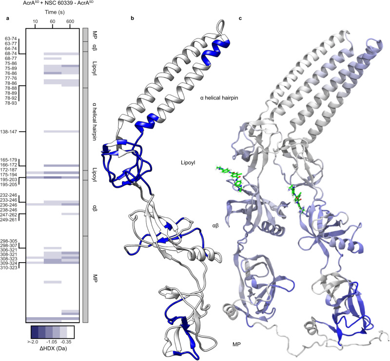
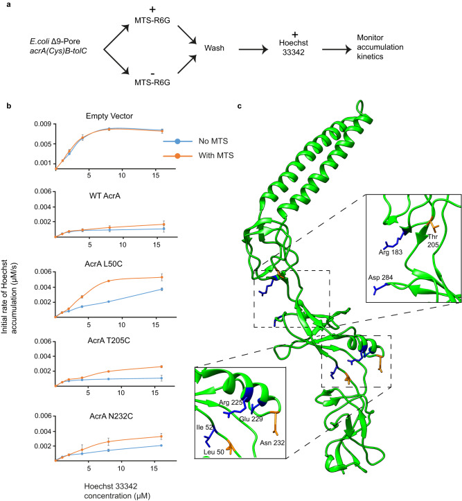
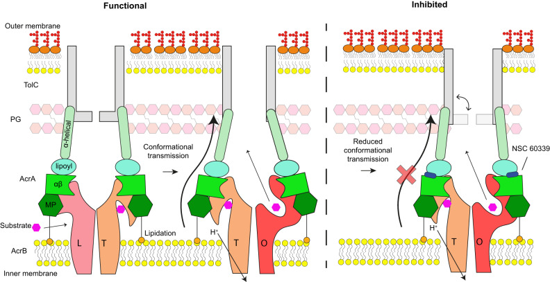
Membrane efflux pumps play a major role in bacterial multidrug resistance. The tripartite multidrug efflux pump system from Escherichia coli, AcrAB-TolC, is a target for inhibition to lessen resistance development and restore antibiotic efficacy, with homologs in other ESKAPE pathogens. Here, we rationalize a mechanism of inhibition against the periplasmic adaptor protein, AcrA, using a combination of hydrogen/deuterium exchange mass spectrometry, cellular efflux assays, and molecular dynamics simulations. We define the structural dynamics of AcrA and find that an inhibitor can inflict long-range stabilisation across all four of its domains, whereas an interacting efflux substrate has minimal effect. Our results support a model where an inhibitor forms a molecular wedge within a cleft between the lipoyl and αβ barrel domains of AcrA, diminishing its conformational transmission of drug-evoked signals from AcrB to TolC. This work provides molecular insights into multidrug adaptor protein function which could be valuable for developing antimicrobial therapeutics.


Conflict of interest statement

The authors declare no competing interests.

Figures

Fig. 1
Fig. 1. AcrA organisation, structure, and constructs.
a Schematic of AcrAB-TolC complex embedded in the cell envelope. LPS Lipopolysaccharide. b Structure of AcrA isolated from the entire AcrAB-TolC complex (PDB:5O66). c Three constructs of AcrA. AcrAL contains Cys25 which is lipidated after signal peptide 1-24 cleavage. AcrAS contains a Cys25Met mutation, and therefore lacks the lipidation and signal peptide. AcrASD contains two AcrAS sequences connected by a TRRIT linker. d Native-MS characterisation of the AcrA constructs at pH 6.0. Proteins buffer was exchanged in 100 mM ammonium acetate buffer prior to MS. AcrAL required the presence of 2× critical micelle concentration (CMC) of DDM at 0.03%. AcrAL presents as a mix of monomers and dimers, AcrAS as monomers and AcrASD as a dimer. Spectra collected on biological replicates and by two different native-MS systems, a time of flight and an orbitrap system, provide confidence these spectral signatures are not artefacts of protein preparation or MS detection. Envelopes marked with an asterisk (*) represent the higher charge state indicative of intrinsic disorder. Masses found in Supplementary Table 1.
Fig. 2
Fig. 2. The effect of NSC 60339 on AcrAS structural dynamics.
a The m/z spectrum for peptide 308-323 under non-deuterating conditions and deuterating conditions with DMSO, NSC 60339 and novobiocin. The centroid is represented by the dotted line, and the mass change of the deuterated samples is written in Daltons. b Chiclet plot displaying the differential HDX (ΔHDX) plots for AcrAS +/− NSC 60339 for all time points collected. Blue signifies areas with decreased HDX between states. We defined significance to be ± ≥ 0.33 Da change (see ‘Methods’ and Supplementary Fig. 9) with a P value ≤ 0.01 in a two-sided Welch’s t test (8 independent measurements: nbiological = 2 and ntechnical = 4). White areas represent regions with insignificant ΔHDX. Source data are provided as a Source data file. c ΔHDX for ((AcrAS + NSC 60339) – AcrAS) for the latest time point is painted onto the AcrA structure (PDB:5O66) using HDeXplosion and Chimera,,. Zoomed in insert of site IV is shown, with the side chains of implicated residues highlighted. d Uptake plots for three peptides in different domains of AcrA. Uptake plots are the average deuterium uptake and error bars indicate the standard deviation (8 independent measurements: nbiological = 2 and ntechnical = 4).
Fig. 3
Fig. 3. MD simulations of NSC 60339 bound to AcrAS.
a Docked structure of NSC 60339 (centre) to AcrA from Darzynkiewicz et al.. F81 and F254 are shown above and below NSC 60339, respectively. b Zoomed-in view of the NSC 60339 binding pocket cleft. AcrA is shown in a surface representation. c NSC 60339 structure; calculated pKa values for the dihydroimidazoline groups are > 9, suggesting that it is dicationic at pH 6.0. d AcrA coloured according to the difference in root-mean-square fluctuations (RMSF) between simulations of the bound and apo states, averaged over four replicas for each. Red indicates that the RMSF is greater in the bound state while blue indicates it’s greater in the apo state (the colour range is from −2 Å, blue, to 2 Å, red as indicated by the colour bar). RMSF was calculated over the last 70 ns of each 100-ns simulation.
Fig. 4
Fig. 4. The effect of NSC 60339 on AcrASD structural dynamics.
a Chiclet plot displaying the differential HDX (ΔHDX) plots for AcrASD +/− NSC 60339 for all time points collected. Blue signifies areas with decreased HDX between states. We defined significance as a ± ≥ 0.35 Da change (see ‘Methods’), with a P value ≤ 0.01 in a two-sided Welch’s t test (7 independent measurements: nbiological = 2 and ntechnical = 3-4). White areas represent regions with insignificant ΔHDX. Source data are provided as a Source data file. b ΔHDX for ((AcrASD + NSC 60339) – AcrASD) for the 10 min time point is painted onto the AcrA structure (PDB:5O66) using HDeXplosion and Chimera,,. c Simulated AcrASD coloured according to the difference in RMSF over the last 70 ns of 100-ns simulations of the bound and apo states, averaged over four replicas for each. Blue indicates that the RMSF is greater in the apo state than the bound state while white indicates they are similar in both states (the colour range is from −2 Å, blue, to 0 Å, white).
Fig. 5
Fig. 5. The effect of Cys-reactive MTS probe on the efficiency of AcrAB-TolC.
a Covalent efflux inhibition assay experimental process. MTS-R6G is the Cys reactive probe and Hoechst 33342 is the fluorescent efflux substrate. b E. coli Δ9-Pore cells producing AcrAB-TolC complex carrying the indicated AcrA variants were split into two aliquots and one of the aliquots was treated with a Cys-reactive probe MTS. After incubation for 15 min at 37 °C, cells were washed and the intracellular accumulation of Hoechst was analysed as described previously. Kinetic data were fitted into a burst-single exponential decay function and the calculated initial rates of Hoechst accumulation (μM/s) are plotted as a function of the externally added concentration of Hoechst. Plots are the mean and error bars indicate standard deviation (n = 3) from independent measurements. Source data provided as Source data file. c AcrA structure (PDB 5O66) with mutated residues highlighted. Blue residues have no effect on efflux and orange residues had an effect.
Fig. 6
Fig. 6. Schematic of AcrAB-TolC inhibition by NSC 60339.
Under normal conditions, as AcrB cycles through its three states of its rotational mechanism (L Loose, T Tight, O Open) the conformational transition information is transmitted through AcrA to TolC, ensuring it is in the ‘open’ state for efflux. AcrA becomes conformationally restricted once NSC 60339 ‘wedges’ between its lipoyl and αβ barrel domains, reducing its ability to transmit the conformational movements of AcrB and, subsequently, inhibiting functional rotation and efflux. Schematic based on in situ structure reported in Chen et al..

References

    1. Sun J, Deng Z, Yan A. Bacterial multidrug efflux pumps: mechanisms, physiology and pharmacological exploitations. Biochem. Biophys. Res. Commun. 2014;453:254–267. doi: 10.1016/j.bbrc.2014.05.090. - DOI - PubMed
    1. Murray CJ, et al. Global burden of bacterial antimicrobial resistance in 2019: a systematic analysis. Lancet. 2022;399:629–655. doi: 10.1016/S0140-6736(21)02724-0. - DOI - PMC - PubMed
    1. Levy SB, Bonnie M. Antibacterial resistance worldwide: causes, challenges and responses. Nat. Med. 2004;10:S122–S129. doi: 10.1038/nm1145. - DOI - PubMed
    1. Nikaido H. Multidrug resistance in bacteria. Annu. Rev. Biochem. 2009;78:119–146. doi: 10.1146/annurev.biochem.78.082907.145923. - DOI - PMC - PubMed
    1. Nikaido H, Zgurskaya HI. AcrAB and related multidrug efflux pumps of Escherichia coli. J. Mol. Microbiol. Biotechnol. 2001;3:215–218. - PubMed

Publication types

MeSH terms

Substances