. 2023 Jul 18;14(1):4317.
doi: 10.1038/s41467-023-40024-3.

Adipose tissue and skeletal muscle wasting precede clinical diagnosis of pancreatic cancer

Affiliations

Adipose tissue and skeletal muscle wasting precede clinical diagnosis of pancreatic cancer

Ana Babic et al. Nat Commun. .

Abstract

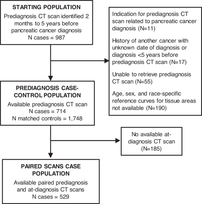
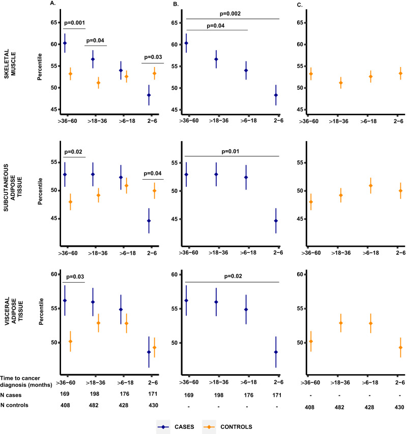
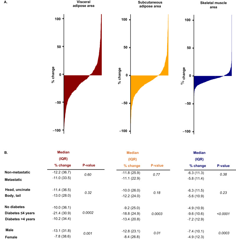
Patients with pancreatic cancer commonly develop weight loss and muscle wasting. Whether adipose tissue and skeletal muscle losses begin before diagnosis and the potential utility of such losses for earlier cancer detection are not well understood. We quantify skeletal muscle and adipose tissue areas from computed tomography (CT) imaging obtained 2 months to 5 years before cancer diagnosis in 714 pancreatic cancer cases and 1748 matched controls. Adipose tissue loss is identified up to 6 months, and skeletal muscle wasting is identified up to 18 months before the clinical diagnosis of pancreatic cancer and is not present in the matched control population. Tissue losses are of similar magnitude in cases diagnosed with localized compared with metastatic disease and are not correlated with at-diagnosis circulating levels of CA19-9. Skeletal muscle wasting occurs in the 1-2 years before pancreatic cancer diagnosis and may signal an upcoming diagnosis of pancreatic cancer.


Conflict of interest statement

M.G.V.H. is a scientific advisor for Agios Pharmaceuticals, iTeos Therapeutics, Sage Therapeutics, Faeth Therapeutics, Droia Ventures, and Auron Therapeutics on topics unrelated to the presented work. B.M.W. received research funding from Celgene, Eli Lilly, Novartis, and Revolution Medicine and consulting fees from Celgene, Grail, Mirati, and Third Rock Ventures unrelated to the presented work. M.B.Y. received research grant from Janssen Pharmaceuticals and fees for peer review services from UpToDate, unrelated to the presented work. The remaining authors declare no competing interests.

Figures

Fig. 1
Fig. 1. Flow diagram of participants from matched pancreatic cancer case-control population.
*Age-, sex-, and race-standardized reference curves for skeletal muscle and adipose tissue areas were previously developed for white non-Hispanic females and males (18–90 years), black females (18–75 years), and black male (18–70 years) participants. Age-, sex-, and race-standardized tissue percentiles could not be calculated from CT scans of participants outside of these categories. CT computed tomography.
Fig. 2
Fig. 2. Comparison of age-, sex-, and race-standardized tissue percentiles between cases and matched controls by time before pancreatic cancer diagnosis.
Data presented as mean values (dots) ± standard errors (bars). A Tissue percentiles in cases and matched controls by time before pancreatic cancer diagnosis. Horizontal lines mark time intervals with measurements significantly different between cases (blue) and controls (yellow) at a P value < 0.05, two-sided Wilcoxon rank-sum test. B, C Tissue percentiles in cases and controls, respectively, by time before pancreatic cancer diagnosis. Horizontal lines mark time intervals with measurements significantly different from the >36 to 60 months interval at a P value < 0.05, Wilcoxon rank-sum test. Source data are provided as a Source Data file.
Fig. 3
Fig. 3. Comparison of body mass index between cases and matched controls by time before pancreatic cancer diagnosis.
Data presented as mean values (dots) ± standard errors (bars). A BMI in cases and matched controls by time before pancreatic cancer diagnosis. Horizontal lines mark time intervals with measurements significantly different between cases (blue) and controls (yellow) at a P value <0.05, two-sided Wilcoxon rank-sum test. B, C BMI in cases and controls, respectively, by time before pancreatic cancer diagnosis. Horizontal lines mark time intervals with measurements significantly different from the >36 to 60 months interval at a P value <0.05, Wilcoxon rank-sum test. Source data are provided as a Source Data file.
Fig. 4
Fig. 4. Change in adipose and skeletal muscle tissue measurements in prediagnostic period in individual patients.
A Waterfall plots of visceral (red), subcutaneous (yellow) and skeletal muscle tissue (blue) measurement changes between prediagnosis and at-diagnosis CT scan. Each vertical line corresponds to one pancreatic cancer case. B Change in adipose and skeletal muscle tissue between prediagnosis and at-diagnosis CT scan by select patient characteristics. Diabetes status and duration related to the time of pancreatic cancer diagnosis. P-values calculated using two-sided Wilcoxon rank sum test. Source data are provided as a Source Data file.

References

    1. Rahib L, et al. Projecting cancer incidence and deaths to 2030: the unexpected burden of thyroid, liver, and pancreas cancers in the United States. Cancer Res. 2014;74:2913–2921. doi: 10.1158/0008-5472.CAN-14-0155. - DOI - PubMed
    1. Siegel RL, Miller KD, Fuchs HE, Jemal A. Cancer statistics, 2021. CA Cancer J. Clin. 2021;71:7–33. doi: 10.3322/caac.21654. - DOI - PubMed
    1. Encarnacion-Rosado J, Kimmelman AC. Harnessing metabolic dependencies in pancreatic cancers. Nat. Rev. Gastroenterol. Hepatol. 2021;18:482–92. doi: 10.1038/s41575-021-00431-7. - DOI - PMC - PubMed
    1. Chari ST, et al. Pancreatic cancer-associated diabetes mellitus: prevalence and temporal association with diagnosis of cancer. Gastroenterology. 2008;134:95–101. doi: 10.1053/j.gastro.2007.10.040. - DOI - PMC - PubMed
    1. Hue JJ, et al. Weight loss as an untapped early detection marker in pancreatic and periampullary cancer. Ann. Surg. Oncol. 2021;28:6283–92. doi: 10.1245/s10434-021-09861-8. - DOI - PubMed

Publication types