. 2024 Feb;53(2):319-328.
doi: 10.1007/s00256-023-04402-8. Epub 2023 Jul 18.

Evaluation of response to neoadjuvant chemotherapy in osteosarcoma using dynamic contrast-enhanced MRI: development and external validation of a model

Affiliations

Evaluation of response to neoadjuvant chemotherapy in osteosarcoma using dynamic contrast-enhanced MRI: development and external validation of a model

Gijsbert M Kalisvaart et al. Skeletal Radiol. 2024 Feb.

Abstract

Objective: To identify which dynamic contrast-enhanced (DCE-)MRI features best predict histological response to neoadjuvant chemotherapy in patients with an osteosarcoma.

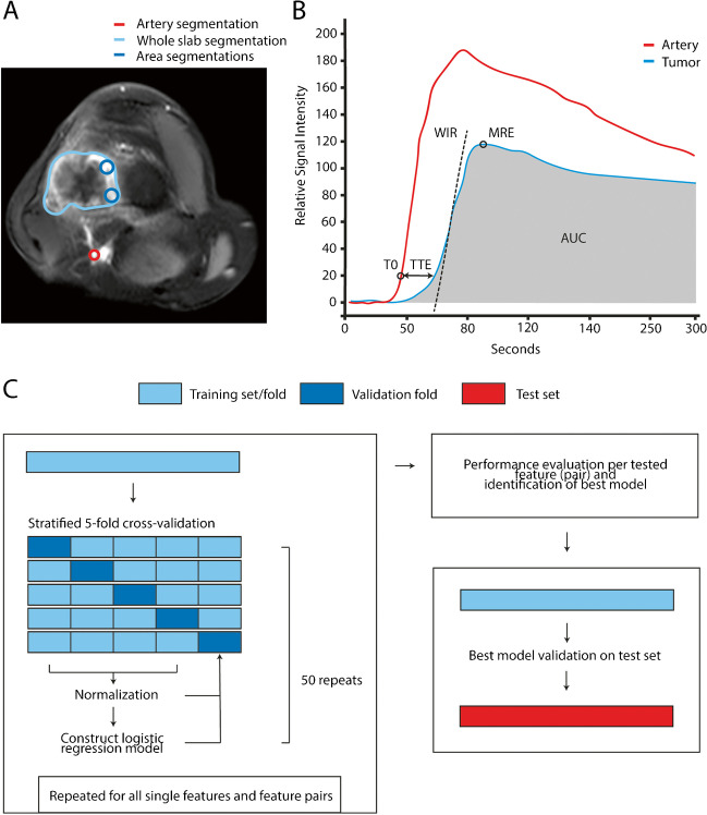
Methods: Patients with osteosarcoma who underwent DCE-MRI before and after neoadjuvant chemotherapy prior to resection were retrospectively included at two different centers. Data from the center with the larger cohort (training cohort) was used to identify which method for region-of-interest selection (whole slab or focal area method) and which change in DCE-MRI features (time to enhancement, wash-in rate, maximum relative enhancement and area under the curve) gave the most accurate prediction of histological response. Models were created using logistic regression and cross-validated. The most accurate model was then externally validated using data from the other center (test cohort).

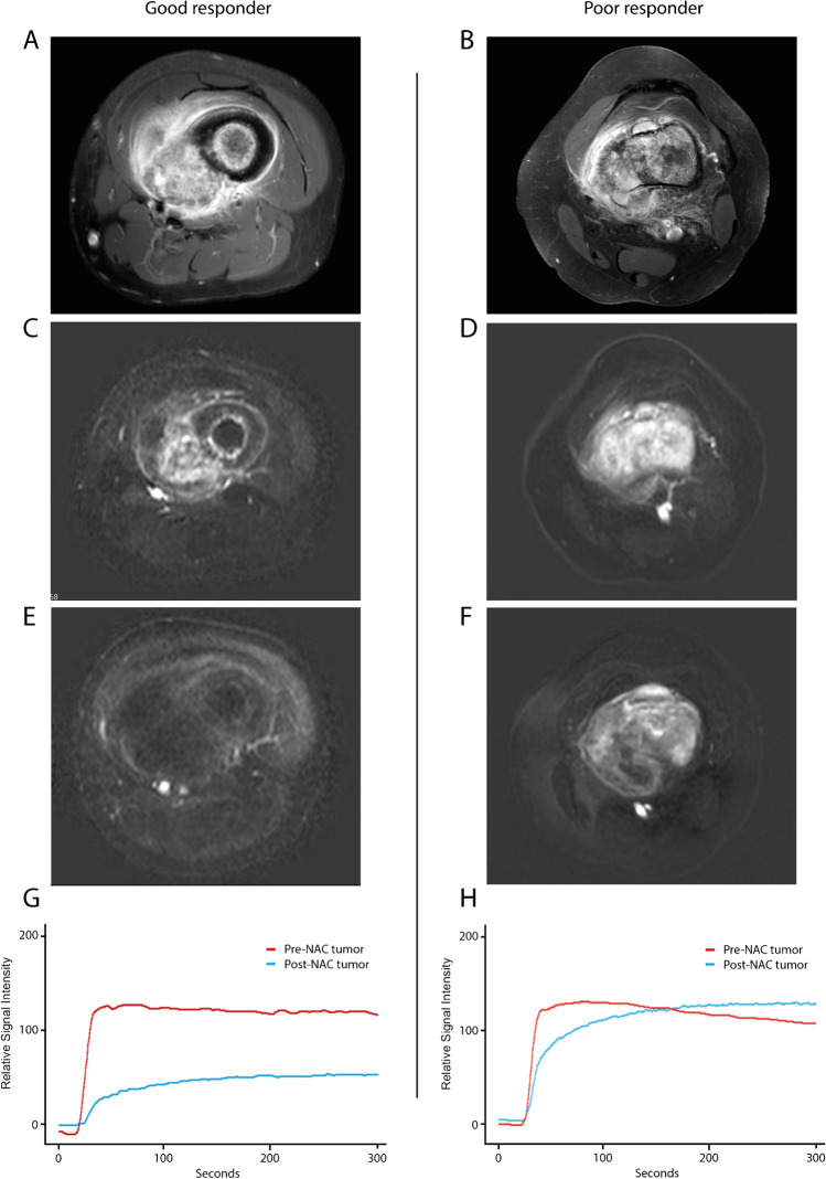
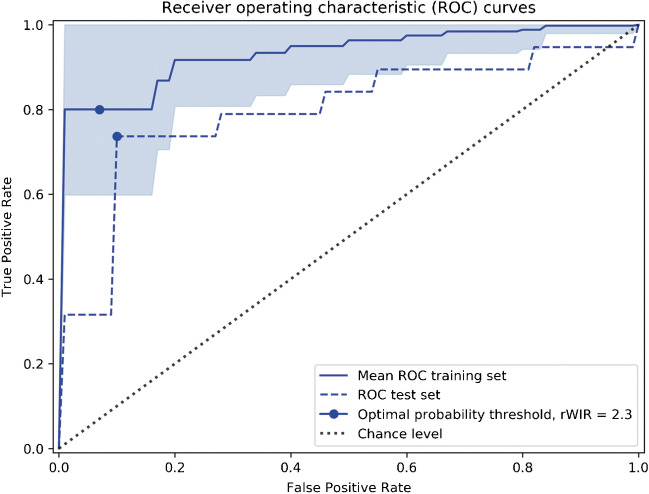
Results: Fifty-five (27 poor response) and 30 (19 poor response) patients were included in training and test cohorts, respectively. Intraclass correlation coefficient of relative DCE-MRI features ranged 0.81-0.97 with the whole slab and 0.57-0.85 with the focal area segmentation method. Poor histological response was best predicted with the whole slab segmentation method using a single feature threshold, relative wash-in rate <2.3. Mean accuracy was 0.85 (95%CI: 0.75-0.95), and area under the receiver operating characteristic curve (AUC-index) was 0.93 (95%CI: 0.86-1.00). In external validation, accuracy and AUC-index were 0.80 and 0.80.

Conclusion: In this study, a relative wash-in rate of <2.3 determined with the whole slab segmentation method predicted histological response to neoadjuvant chemotherapy in osteosarcoma. Consistent performance was observed in an external test cohort.

Keywords: Dynamic contrast-enhanced MRI; External validation; Histological response; Neoadjuvant chemotherapy; Osteosarcoma; Response monitoring.


Conflict of interest statement

G.M. Kalisvaart was the recipient of an educational grant from Philips Electronics Nederland B. V, Eindhoven, The Netherlands, during writing of this manuscript (number: LEI-05). Furthermore, the research presented in the manuscript was supported by a public grant from Health~Holland TKI Life Sciences & Health (number: LSHM18089). Other authors had nothing to disclose. The authors declare that they have no conflict of interest.

Figures

Fig. 1
Fig. 1
Overview of segmentation methods, feature extraction, model building and model testing. A; Dynamic contrast-enhanced MRI image of an osteosarcoma in the distal femur showing the 2D segmentation of the entire tumor (whole slab, light blue area) and regions-of-interest placed in the most intensely and early enhancing parts of the tumor (focal area method, dark blue circles). Arterial segmentation (red circle) is performed as reference tissue comparison to the tumoral segmentations. B; Schematic dynamic contrast-enhanced MRI derived time-intensity curve and perfusion features for an artery and a tumor region depicting changes in average pixel signal intensity over time due to influx and outflux of gadolinium contrast medium and its distribution over the vascular versus tumoral compartments. The features time to enhancement (TTE), wash-in rate (WIR), maximum relative enhancement (MRE) and area under the curve (AUC) are illustrated. C; Overview of model training with logistic regression, internal cross-validation, model selection and external validation on a test cohort. Models were cross-validated to test all single features separately and all feature pairs per segmentation method. AUC area under the curve, MRE maximum relative enhancement, T0 time of onset of enhancement, TTE time to enhancement, WIR wash-in rate
Fig. 2
Fig. 2
Differences in (dynamic) contrast-enhanced MRI images and time-intensity curve analysis in osteosarcoma patients between good and poor responders to neoadjuvant chemotherapy. A–B; Pre-neoadjuvant chemotherapy T1-weighted gadolinium contrast-enhanced spectral pre-saturation with inversion recovery (SPIR) images of an osteosarcoma in the femur diaphysis and in the distal femur in a good (A) and poor (B) responder, respectively. C–D; Subtraction images of the pre-neoadjuvant chemotherapy dynamic contrast-enhanced MRI sequence at 6 s after arrival of gadolinium contrast medium in a good (C) and poor (D) responder. E–F; Subtraction images of the post-neoadjuvant chemotherapy dynamic contrast-enhanced MRI sequence at 6 s after arrival of gadolinium contrast medium showing an absence of residual enhancement in the good responder (E) and persistent and heterogeneous enhancement of the poor responder (F). After resection, the percentage of viable tumor cells post-neoadjuvant chemotherapy was estimated to be 1% versus 20–40% in the good and poor responder, respectively. G–H; Dynamic contrast-enhanced MRI-derived time-intensity curves in corresponding tumors (whole slab method) pre-neoadjuvant chemotherapy (red) and post-neoadjuvant chemotherapy (blue) in a good responder (G) and in a poor responder (H). NAC neoadjuvant chemotherapy
Fig. 3
Fig. 3
Representative histology of osteosarcomas depicted in Fig. 2 (good versus poor responder). Black scale bars represent 100 μm. A–B; Good responder post-neoadjuvant chemotherapy with 1% vital tumor cells remaining and showing loose edematous as well as more condensed fibrotic areas with remnants of tumor osteoid (arrows). C–D; Poor responder post-neoadjuvant chemotherapy with 30% vital tumor cells remaining and showing pre-existing lamellar bone, surrounded by a proliferation of pleomorphic and vital tumor cells depositing osteoid
Fig. 4
Fig. 4
Receiver operating characteristic curves for the final model (rWIR as a single feature of the whole slab segmentation method) with internal cross-validation and external validation on the test cohort results. ROC receiver operating characteristic, rWIR relative wash-in rate

References

    1. Bielack SS, Kempf-Bielack B, Delling G, et al. Prognostic factors in high-grade osteosarcoma of the extremities or trunk: an analysis of 1,702 patients treated on neoadjuvant cooperative osteosarcoma study group protocols. J Clin Oncol. 2002;20(3):776–790. doi: 10.1200/JCO.2002.20.3.776. - DOI - PubMed
    1. Huvos AG, Rosen G, Marcove RC. Primary osteogenic sarcoma: pathologic aspects in 20 patients after treatment with chemotherapy en bloc resection, and prosthetic bone replacement. Arch Pathol Lab Med. 1977;101(1):14–18. - PubMed
    1. Guo J, Reddick WE, Glass JO, et al. Dynamic contrast-enhanced magnetic resonance imaging as a prognostic factor in predicting event-free and overall survival in pediatric patients with osteosarcoma. Cancer. 2012;118(15):3776–3785. doi: 10.1002/cncr.26701. - DOI - PMC - PubMed
    1. Byun BH, Kong CB, Lim I, et al. Combination of 18F-FDG PET/CT and diffusion-weighted MR imaging as a predictor of histologic response to neoadjuvant chemotherapy: preliminary results in osteosarcoma. J Nucl Med. 2013;54(7):1053–1059. doi: 10.2967/jnumed.112.115964. - DOI - PubMed
    1. Habre C, Dabadie A, Loundou AD, et al. Diffusion-weighted imaging in differentiating mid-course responders to chemotherapy for long-bone osteosarcoma compared to the histologic response: an update. Pediatr Radiol. 2021;51(9):1714–1723. doi: 10.1007/s00247-021-05037-4. - DOI - PMC - PubMed

MeSH terms

Grants and funding