

Stimulation parameters for directional vagus nerve stimulation
- PMID: 37464423
- PMCID: PMC10353120
- DOI: 10.1186/s42234-023-00117-2
Stimulation parameters for directional vagus nerve stimulation
Abstract
Background: Autonomic nerve stimulation is used as a treatment for a growing number of diseases. We have previously demonstrated that application of efferent vagus nerve stimulation (eVNS) has promising glucose lowering effects in a rat model of type 2 diabetes. This paradigm combines high frequency pulsatile stimulation to block nerve activation in the afferent direction with low frequency stimulation to activate the efferent nerve section. In this study we explored the effects of the parameters for nerve blocking on the ability to inhibit nerve activation in the afferent direction. The overarching aim is to establish a blocking stimulation strategy that could be applied using commercially available implantable pulse generators used in the clinic.
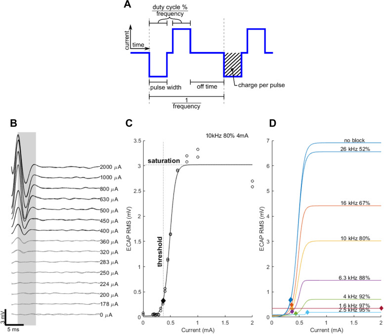
Methods: Male rats (n = 20) had the anterior abdominal vagus nerve implanted with a multi-electrode cuff. Evoked compound action potentials (ECAP) were recorded at the proximal end of the electrode cuff. The efficacy of high frequency stimulation to block the afferent ECAP was assessed by changes in the threshold and saturation level of the response. Blocking frequency and duty cycle of the blocking pulses were varied while maintaining a constant 4 mA current amplitude.
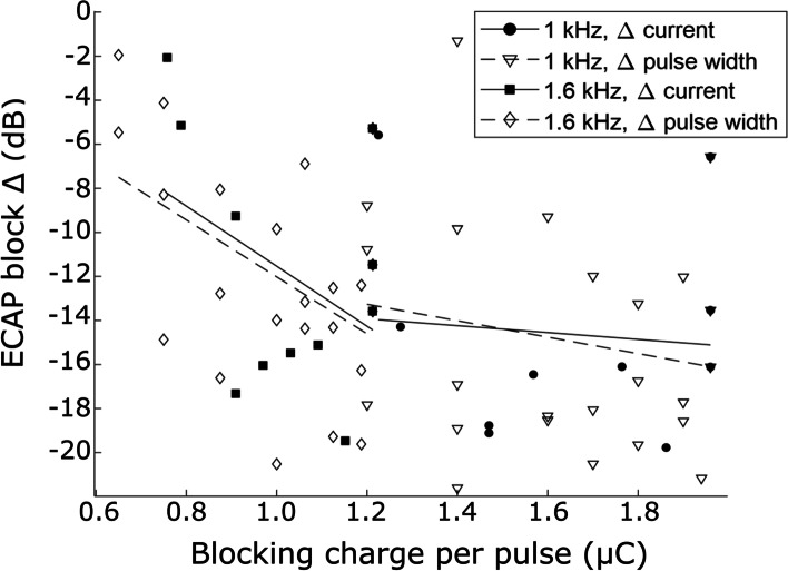
Results: During application of blocking at lower frequencies (≤ 4 kHz), the ECAP threshold increased (ANOVA, p < 0.001) and saturation level decreased (p < 0.001). Application of higher duty cycles (> 70%) led to an increase in evoked neural response threshold (p < 0.001) and a decrease in saturation level (p < 0.001). During the application of a constant pulse width and frequency (1 or 1.6 kHz, > 70% duty cycle), the charge delivered per pulse had a significant influence on the magnitude of the block (ANOVA, p = 0.003), and was focal (< 2 mm range).
Conclusions: This study has determined the range of frequencies, duty cycles and currents of high frequency stimulation that generate an efficacious, focal axonal block of a predominantly C-fiber tract. These findings could have potential application for the treatment of type 2 diabetes.
Keywords: Bioelectric medicine; Medical devices; Metabolic disease; Nerve blocking; Peripheral nerve stimulation.
© 2023. The Author(s).
Conflict of interest statement
JBF is on the
Figures


Similar articles
-
Blood glucose modulation and safety of efferent vagus nerve stimulation in a type 2 diabetic rat model.Physiol Rep. 2022 Apr;10(8):e15257. doi: 10.14814/phy2.15257. Physiol Rep. 2022. PMID: 35439355 Free PMC article.
-
In vivo peripheral nerve activation using sinusoidal low-frequency alternating currents.Artif Organs. 2022 Oct;46(10):2055-2065. doi: 10.1111/aor.14347. Epub 2022 Jun 30. Artif Organs. 2022. PMID: 35730955 Free PMC article.
-
kHz-frequency electrical stimulation selectively activates small, unmyelinated vagus afferents.Brain Stimul. 2022 Nov-Dec;15(6):1389-1404. doi: 10.1016/j.brs.2022.09.015. Epub 2022 Oct 11. Brain Stimul. 2022. PMID: 36241025 Free PMC article.
-
Electrically evoked compound action potential (ECAP) of the cochlear nerve in response to pulsatile electrical stimulation of the cochlea in the rat: effects of stimulation at high rates.Audiology. 1998 Nov-Dec;37(6):353-71. doi: 10.3109/00206099809072989. Audiology. 1998. PMID: 9888192
-
A Respiratory Marker Derived From Left Vagus Nerve Signals Recorded With Implantable Cuff Electrodes.Neuromodulation. 2018 Apr;21(3):269-275. doi: 10.1111/ner.12630. Epub 2017 Jul 11. Neuromodulation. 2018. PMID: 28699322 Review.
Cited by
-
Importance of cardiac-synchronized vagus nerve stimulation parameters on the provoked chronotropic response for different levels of cardiac innervation.Front Physiol. 2024 May 21;15:1379936. doi: 10.3389/fphys.2024.1379936. eCollection 2024. Front Physiol. 2024. PMID: 38835728 Free PMC article.
-
Therapeutic Effects of Electroencephalogram-Based Bioelectric Stimulation on Cognitive-Behavioural Outcomes in Children With Dual Diagnosis of Autism Spectrum Disorder and Intellectual Disability.Actas Esp Psiquiatr. 2025 Aug;53(4):802-812. doi: 10.62641/aep.v53i4.1975. Actas Esp Psiquiatr. 2025. PMID: 40791035 Free PMC article.
-
Selective efferent vagal stimulation in heart failure.Exp Physiol. 2024 Dec;109(12):2001-2005. doi: 10.1113/EP090866. Epub 2023 Sep 26. Exp Physiol. 2024. PMID: 37755233 Free PMC article. Review.
References
-
- Ahren B, Paquette TL, Taborsky GJ., Jr Effect and mechanism of vagal nerve stimulation on somatostatin secretion in dogs. Am J Physiol. 1986;250:E212–217. - PubMed
Grants and funding
LinkOut - more resources
Full Text Sources