

Intraductal papillary mucinous neoplasm of the biliary tract in the caudate lobe of the liver: a case report and literature review
- PMID: 37465111
- PMCID: PMC10351580
- DOI: 10.3389/fonc.2023.1114514
Intraductal papillary mucinous neoplasm of the biliary tract in the caudate lobe of the liver: a case report and literature review
Abstract
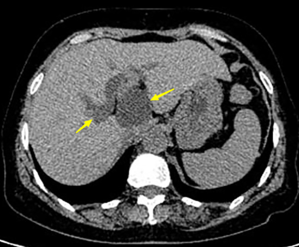
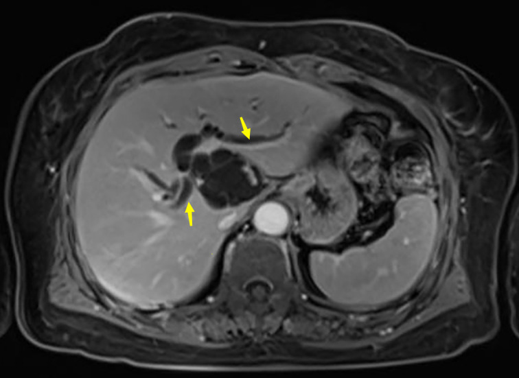
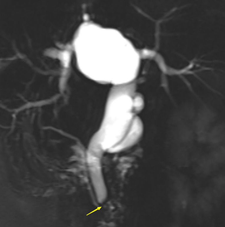
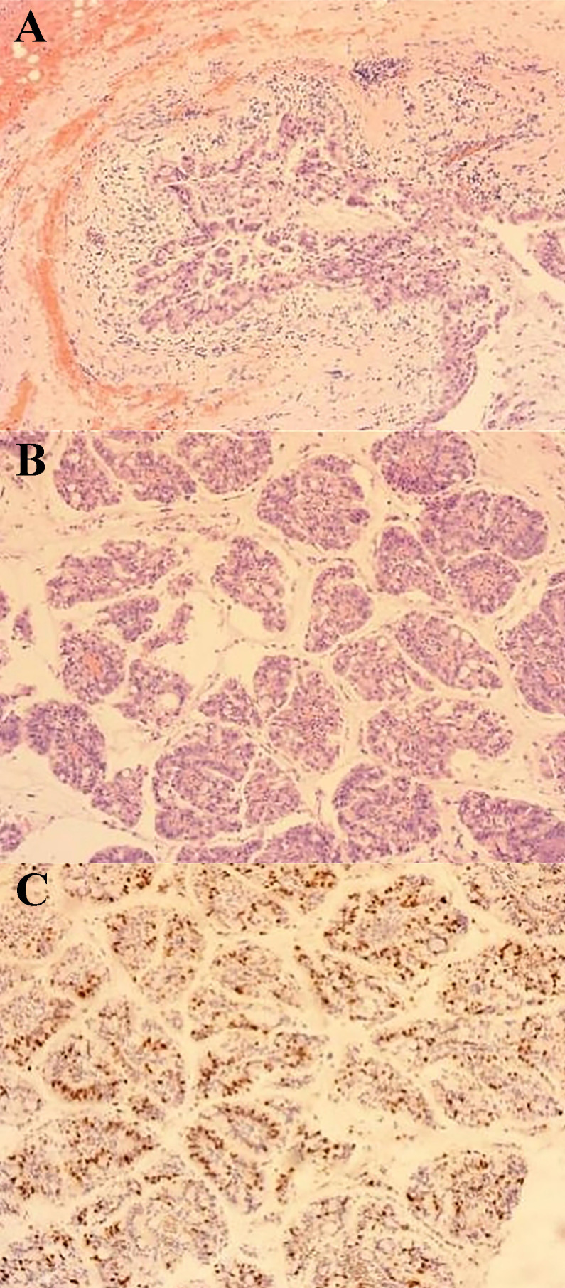
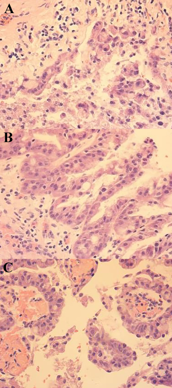
An intraductal papillary mucinous neoplasm of the biliary tract (BT-IPMN) in the caudate lobe of the liver is a rare tumor originating from the bile duct. Approximately 40% of the intraductal papillary neoplasms of the biliary tract (IPNB) secrete mucus and can grow in the intrahepatic or extrahepatic bile ducts. A 65-year-old woman presented with recurrent episodes of right upper pain. She developed her first episode 8 years ago, which resolved spontaneously. The frequency of symptoms has increased in the last 2 years. She underwent laparoscopic hepatectomy and choledochal exploration and was pathologically diagnosed with a rare BT-IPMN of the caudate lobe after admission. Here, we review studies on IPNB cases and systematically describe the pathological type, diagnosis, and treatment of IPNB to provide a valuable reference for hepatobiliary surgeons in the diagnosis and treatment of this disease.
Keywords: bile duct; biliary tract surgery; diagnosis; intraductal papillary mucinous neoplasm; pathology; treatment.
Copyright © 2023 Zhu, Ni, Wang, Ma, Yang, Gao, Zhu, Zhou, Chang, Lu and Liu.
Conflict of interest statement
The authors declare that the research was conducted in the absence of any commercial or financial relationships that could be construed as a potential conflict of interest.
Figures
Similar articles
-
Intraductal Papillary Mucinous Neoplasm of the Bile Duct: A Case Report.Cureus. 2025 Feb 8;17(2):e78749. doi: 10.7759/cureus.78749. eCollection 2025 Feb. Cureus. 2025. PMID: 40070623 Free PMC article.
-
Long-term observation and treatment of a widespread intraductal papillary neoplasm of the bile duct extending from the intrapancreatic bile duct to the bilateral intrahepatic bile duct: A case report.Int J Surg Case Rep. 2017;38:166-171. doi: 10.1016/j.ijscr.2017.07.031. Epub 2017 Jul 23. Int J Surg Case Rep. 2017. PMID: 28763696 Free PMC article.
-
Clinicopathological characterization of so-called "cholangiocarcinoma with intraductal papillary growth" with respect to "intraductal papillary neoplasm of bile duct (IPNB)".Int J Clin Exp Pathol. 2014 May 15;7(6):3112-22. eCollection 2014. Int J Clin Exp Pathol. 2014. PMID: 25031730 Free PMC article.
-
Current status of diagnosis and therapy for intraductal papillary neoplasm of the bile duct.World J Gastroenterol. 2021 Apr 21;27(15):1569-1577. doi: 10.3748/wjg.v27.i15.1569. World J Gastroenterol. 2021. PMID: 33958844 Free PMC article. Review.
-
Intraductal papillary neoplasm of the bile ducts: A case report and literature review.World J Gastroenterol. 2015 Nov 21;21(43):12498-504. doi: 10.3748/wjg.v21.i43.12498. World J Gastroenterol. 2015. PMID: 26604656 Free PMC article. Review.
Cited by
-
Pancreatic neuroendocrine neoplasms coexisting with biliary intraductal papillary mucinous neoplasm: A case report and review of literature.World J Gastrointest Oncol. 2025 Apr 15;17(4):100497. doi: 10.4251/wjgo.v17.i4.100497. World J Gastrointest Oncol. 2025. PMID: 40235901 Free PMC article.
References
Publication types
LinkOut - more resources
Full Text Sources