

Single cell analysis of transcriptome and open chromatin reveals the dynamics of hair follicle stem cell aging
- PMID: 37465120
- PMCID: PMC10350644
- DOI: 10.3389/fragi.2023.1192149
Single cell analysis of transcriptome and open chromatin reveals the dynamics of hair follicle stem cell aging
Abstract
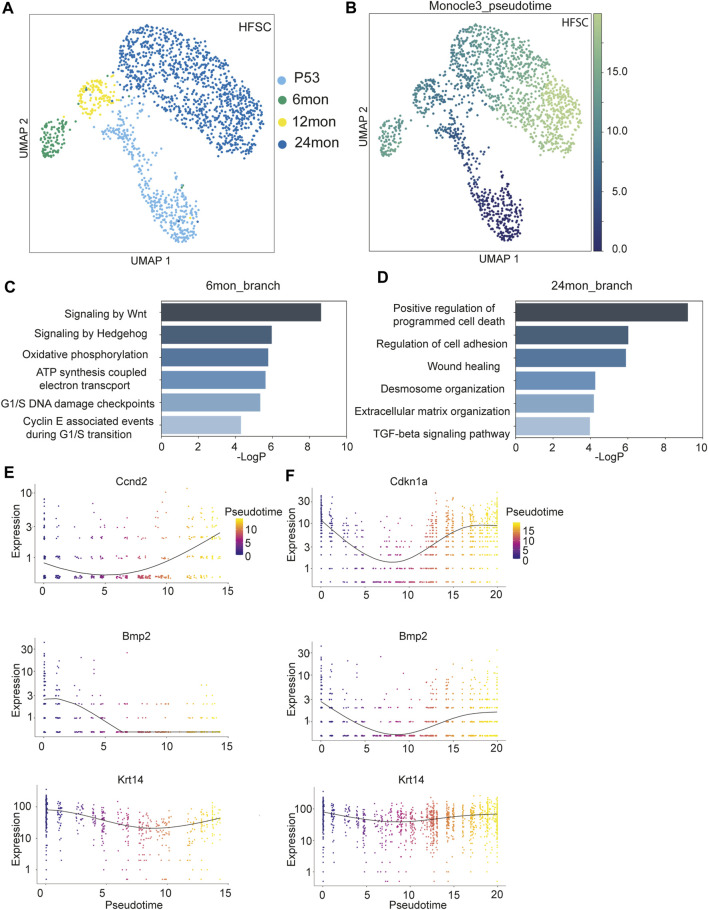
Aging is defined as the functional decline of tissues and organisms, leading to many human conditions, such as cancer, neurodegenerative diseases, and hair loss. Although stem cell exhaustion is widely recognized as a hallmark of aging, our understanding of cell state changes-specifically, the dynamics of the transcriptome and open chromatin landscape, and their relationship with aging-remains incomplete. Here we present a longitudinal, single-cell atlas of the transcriptome and open chromatin landscape for epithelia cells of the skin across various hair cycle stages and ages in mice. Our findings reveal fluctuating hair follicle stem cell (HF-SC) states, some of which are associated with the progression of the hair cycle during aging. Conversely, inner bulge niche cells display a more linear progression, seemingly less affected by the hair cycle. Further analysis of the open chromatin landscape, determined by single-cell Assay for Transposase-Accessible Chromatin (ATAC) sequencing, demonstrates that reduced open chromatin regions in HF-SCs are associated with differentiation, whereas gained open chromatin regions in HF-SCs are linked to the transcriptional control of quiescence. These findings enhance our understanding of the transcriptional dynamics in HF-SC aging and lay the molecular groundwork for investigating and potentially reversing the aging process in future experimental studies.
Keywords: cell adhesion; hair follicle stem cell; quiescence; scATACseq; scRNAseq; single cell genomics; stem cell exhaustion.
Copyright © 2023 Zhang, Wang, Dowell and Yi.
Conflict of interest statement
The authors declare that the research was conducted in the absence of any commercial or financial relationships that could be construed as a potential conflict of interest.
Figures


Similar articles
-
Escape of hair follicle stem cells causes stem cell exhaustion during aging.Nat Aging. 2021 Oct;1(10):889-903. doi: 10.1038/s43587-021-00103-w. Epub 2021 Oct 4. Nat Aging. 2021. PMID: 37118327 Free PMC article.
-
FOXC1 maintains the hair follicle stem cell niche and governs stem cell quiescence to preserve long-term tissue-regenerating potential.Proc Natl Acad Sci U S A. 2016 Mar 15;113(11):E1506-15. doi: 10.1073/pnas.1601569113. Epub 2016 Feb 24. Proc Natl Acad Sci U S A. 2016. PMID: 26912458 Free PMC article.
-
High-resolution single-cell transcriptomics reveals heterogeneity of self-renewing hair follicle stem cells.Exp Dermatol. 2021 Apr;30(4):457-471. doi: 10.1111/exd.14262. Epub 2021 Jan 6. Exp Dermatol. 2021. PMID: 33319418 Free PMC article.
-
Advances in a rapidly emerging field of hair follicle stem cell research.Coll Antropol. 2014 Mar;38(1):373-8. Coll Antropol. 2014. PMID: 24851645 Review.
-
Aging in hair follicle stem cells and niche microenvironment.J Dermatol. 2017 Oct;44(10):1097-1104. doi: 10.1111/1346-8138.13897. Epub 2017 Jun 8. J Dermatol. 2017. PMID: 28593683 Review.
Cited by
-
Reprogramming of epidermal keratinocytes by PITX1 transforms the cutaneous cellular landscape and promotes wound healing.JCI Insight. 2024 Dec 20;9(24):e182844. doi: 10.1172/jci.insight.182844. JCI Insight. 2024. PMID: 39480496 Free PMC article.
-
PIEZO1-mediated calcium signaling reinforces mechanical properties of hair follicle stem cells to promote quiescence.Sci Adv. 2025 May 30;11(22):eadt2771. doi: 10.1126/sciadv.adt2771. Epub 2025 May 28. Sci Adv. 2025. PMID: 40435254 Free PMC article.
-
Determinants of Chromatin Organization in Aging and Cancer-Emerging Opportunities for Epigenetic Therapies and AI Technology.Genes (Basel). 2024 May 29;15(6):710. doi: 10.3390/genes15060710. Genes (Basel). 2024. PMID: 38927646 Free PMC article. Review.
-
CXCL12 Drives Reversible Fibroimmune Remodeling in Androgenetic Alopecia Revealed by Single-Cell RNA Sequencing.Int J Mol Sci. 2025 Jul 8;26(14):6568. doi: 10.3390/ijms26146568. Int J Mol Sci. 2025. PMID: 40724819 Free PMC article.
-
SLC6A6-Mediated Taurine Uptake Sustains Corneal Epithelial Stem/Progenitor Cell Function to Counteract Age-Related Dysfunction.Invest Ophthalmol Vis Sci. 2025 Jun 2;66(6):25. doi: 10.1167/iovs.66.6.25. Invest Ophthalmol Vis Sci. 2025. PMID: 40478558 Free PMC article.
References
Grants and funding
LinkOut - more resources
Full Text Sources
Molecular Biology Databases
Research Materials
Miscellaneous