

Characterization of peripheral blood inflammatory indicators and OCT imaging biological markers in diabetic retinopathy with or without nephropathy
- PMID: 37465123
- PMCID: PMC10351984
- DOI: 10.3389/fendo.2023.1160615
Characterization of peripheral blood inflammatory indicators and OCT imaging biological markers in diabetic retinopathy with or without nephropathy
Abstract
Objective: To observe the distribution characteristics of peripheral blood inflammatory indexes and retinal macular area optical coherence tomography (OCT) imaging biomarkers in patients with diabetic retinopathy (DR) with or without diabetic nephropathy (DN), in order to seek clinical biomarkers that can predict the development of DR and DN.
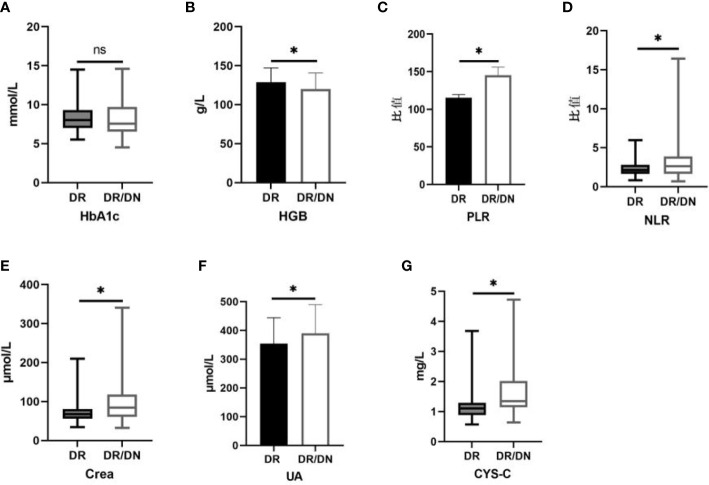
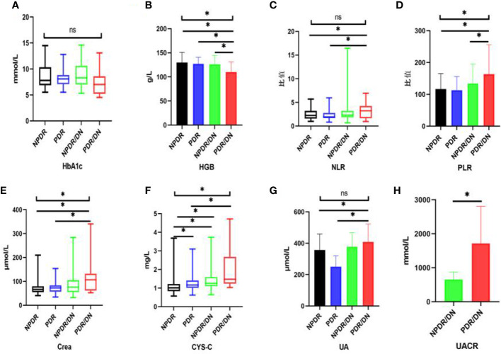
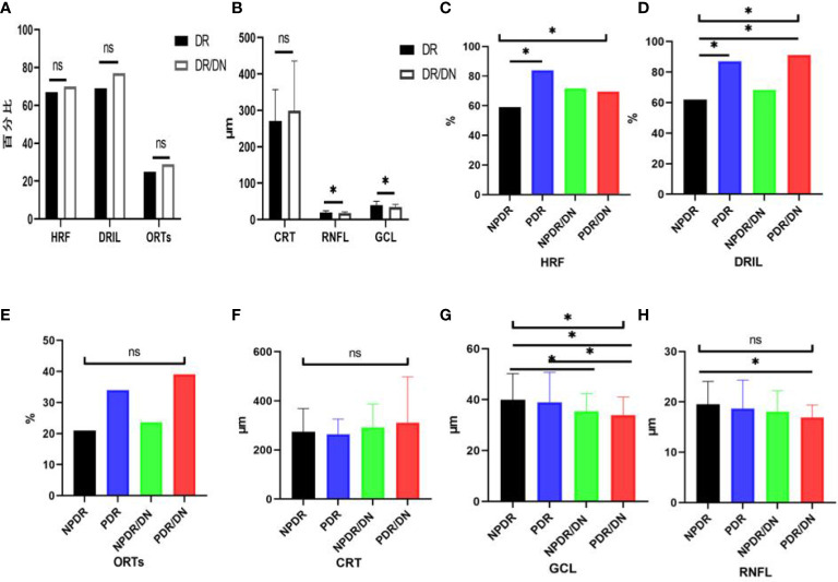
Methods: A total of 169 inpatients with DR who visited the ophthalmology department of the Affiliated Hospital of Chengdu University of Traditional Chinese Medicine from October 2020 to June 2022 and had complete clinical data were collected, and the patients with DR were divided into two major groups, DR and DR/DN, according to whether they had DN, and then further divided into four subgroups, Non-proliferative DR(NPDR), proliferative DR(PDR), NPDR/DN and PDR/DN, according to the stage of DR. The distribution characteristics of peripheral blood inflammatory indexes [Neutrophil to lymphocyte ratio(NLR) and Platelet to neutrophil ratio(PLR)], renal function indexes [Cystatin-C(CYS-C), Creatinine(Crea), Uric acid(UA)and Urinary albumin to creatinine ratio(UACR)] and OCT imaging indexes [Hyperreflective foci(HRF), Disorgnization of retinal inner layers(DRIL), Outer retinal tubulations(ORTs), Central retinal thickness(CRT), Retinal nerve fiber layer(RNFL) and Ganglion cell layer(GCL)] were analyzed between the above subgroups.
Results: There was no difference between DR and DR/DN groups in terms of gender, family history of diabetes, duration of diabetes and Body mass index(BMI) (P>0.05), the mean age of the DR/DN group was significantly lower than that of the DR group (P<0.05), and the proportion of the DR/DN group with a history of hypertension was significantly higher than that of the DR group (P<0.05); there was no significant difference in hemoglobin A1C(HbA1c) between DR and DR/DN groups (P>0.05). (P>0.05), Hemoglobin(HGB) was significantly higher in the DR group than in the DR/DN group (P <0.05), NLR, PLR, Crea, UA and CYS-C were significantly higher in the DR/DN group than in the DR group (P<0.05); there was no significant difference in the comparison of HRF, DRIL, ORTs positive rate and CRT between the DR and DR/DN groups (P>0.05). RNFL and GCL thickness were significantly lower in the DR/DN group than in the DR group (P<0.05); history of hypertension (OR=2.759), NLR (OR=1.316), PLR (OR=1.009), Crea (OR=1.018), UA (OR=1.004), CYS-C (OR=3.742) were the independent (OR=0.951), age (OR=0.951), HGB (OR=0.976), RNFL (OR=0.909) and GCL (OR=0.945) were independent protective factors for DR/DN; RNFL (OR=0.899) and GCL (OR=0.935) were independent protective factors for NPDR/DN, RNFL (OR=0.852) and GCL (OR=0.928) were independent protective factors for PDR/DN. ROC curve analysis showed that the area under the curve (AUC) for CYS-C, PLR, Crea, UA and the combination of the four indicators to predict DR/DN were 0.717, 0.625, 0.647, 0.616 and 0.717, respectively.
Conclusions: (1) Low age combined with hypertension HGB, NLR, PLR, CYS-C, Crea and UA may be serum biological markers for predicting DN in DR; meanwhile, PLR, CYS-C, Crea, UA and the combination of the four indicators can be used for risk assessment and adjunctive diagnosis of DN in DR combined with hypertension. (2) The RNFL and GCL thickness in the temporal aspect of the central macular sulcus may be imaging biological markers for predicting DN in DR; meanwhile, GCL thickness may have important value for risk prediction and diagnosis of DN in combination with DR.
Keywords: diabetic nephropathy; diabetic retinopathy; inflammation; neutrophil-to-lymphocyte ratio (NLR); optical coherence tomography (OCT); platelet-to-lymphocyte ratio (PLR).
Copyright © 2023 Xiaodong, Xuejun, Xiaojuan, Yu and Mingchao.
Conflict of interest statement
The authors declare that the research was conducted in the absence of any commercial or financial relationships that could be construed as a potential conflict of interest.
Figures
Similar articles
-
Exploring the correlations between six serological inflammatory markers and different stages of type 2 diabetic retinopathy.Sci Rep. 2025 Jan 10;15(1):1567. doi: 10.1038/s41598-025-85164-2. Sci Rep. 2025. PMID: 39794420 Free PMC article.
-
Disorganization of retinal inner layers correlates with ellipsoid zone disruption and retinal nerve fiber layer thinning in diabetic retinopathy.J Diabetes Complications. 2019 Aug;33(8):550-553. doi: 10.1016/j.jdiacomp.2019.05.006. Epub 2019 May 17. J Diabetes Complications. 2019. PMID: 31186163
-
Optical Coherence Tomography-Based Grading of Diabetic Macular Edema Is Associated with Systemic Inflammatory Indices and Imaging Biomarkers.Ophthalmic Res. 2024;67(1):96-106. doi: 10.1159/000535199. Epub 2024 Jan 11. Ophthalmic Res. 2024. PMID: 38211574
-
Uric Acid and Diabetic Retinopathy: A Systematic Review and Meta-Analysis.Front Public Health. 2022 May 31;10:906760. doi: 10.3389/fpubh.2022.906760. eCollection 2022. Front Public Health. 2022. PMID: 35712295 Free PMC article.
-
Vascular Changes and Neurodegeneration in the Early Stages of Diabetic Retinopathy: Which Comes First?Ophthalmic Res. 2016;56(1):1-9. doi: 10.1159/000444498. Epub 2016 Apr 2. Ophthalmic Res. 2016. PMID: 27035578 Review.
Cited by
-
Exploring leukocyte differential count ratio profiles as inflammatory biomarkers in diabetic retinopathy: a systematic review and meta-analysis.BMC Ophthalmol. 2025 May 1;25(1):265. doi: 10.1186/s12886-025-04075-y. BMC Ophthalmol. 2025. PMID: 40312699 Free PMC article.
References
-
- Moriya T, Matsubara M, Kishihara E, Yoshida Y, Ouchi M. Type 2 diabetic patients with diabetic retinopathy and concomitant microalbuminuria showed typical diabetic glomerulosclerosis and progressive renal dysfunction. J Diabetes Complications (2016) 6:1111–6. doi: 10.1016/j.jdiacomp.2016.04.007 - DOI - PubMed
Publication types
MeSH terms
Substances
LinkOut - more resources
Full Text Sources
Medical
Research Materials