. 2023 Jul 14:11:e15727.
doi: 10.7717/peerj.15727. eCollection 2023.

IL-17A exacerbates psoriasis in a STAT3 overexpressing mouse model

Affiliations

IL-17A exacerbates psoriasis in a STAT3 overexpressing mouse model

Xinran Xie et al. PeerJ. .

Abstract

Background: Psoriasis is an autoimmune skin disease characterized by immunocyte activation, excessive proliferation, and abnormal differentiation of keratinocytes. Signal transducers and activators of transcription 3 (STAT3) play a crucial role in linking activated keratinocytes and immunocytes during psoriasis development. T helper (Th) 17 cells and secreted interleukin (IL)-17A contribute to its pathogenesis. IL-17A treated STAT3 overexpressing mouse model might serve as an animal model for psoriasis.

Methods: In this study, we established a mouse model of psoriasiform dermatitis by intradermal IL-17A injection in STAT3 overexpressing mice. Transcriptome analyses were performed on the skin of wild type (WT), STAT3, and IL-17A treated STAT3 mice. Bioinformatics-based functional enrichment analysis was conducted to predict biological pathways. Meanwhile, the morphological and pathological features of skin lesions were observed, and the DEGs were verified by qPCR.

Results: IL-17A treated STAT3 mice skin lesions displayed the pathological features of hyperkeratosis and parakeratosis. The DEGs between IL-17A treated STAT3 mice and WT mice were highly consistent with those observed in psoriasis patients, including S100A8, S100A9, Sprr2, and LCE. Gene ontology (GO) analysis of the core DEGs revealed a robust immune response, chemotaxis, and cornified envelope, et al. The major KEGG enrichment pathways included IL-17 and Toll-like receptor signaling pathways.

Conclusion: IL-17A exacerbates psoriasis dermatitis in a STAT3 overexpressing mouse.

Keywords: Bioinformatics; IL-17A; Mouse model; Psoriasis; STAT3.


Conflict of interest statement

The authors declare that they have no competing interests.

Figures
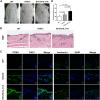
Figure 1
Figure 1. Skin phenotypes in WT, STAT3, and IL-17A treated STAT3 mice.
STAT3 mice and IL-17A treated STAT3 mice vs. wild type (C57BL/6) mice, respectively (n = 10). (A) Images of psoriasiform skin. (B) Skin biopsies were stained with hematoxylin and eosin (H&E). Mitotic basal cells are shown in the insets. Scale bar = 200 μm. Representative PCNA and involucrin immunostained images of the dorsal skin. Scale bar = 50 μm. (C) Epidermal thickness was measured using the image analysis system. Differences were considered statistically significant at *P < 0.05, **P < 0.01, and ***P < 0.001.
Figure 2
Figure 2. Differential gene expression in WT, STAT3, and IL-17A treated STAT3 mice.
(A) Hierarchical clustering of gene expressions from the skin of WT, STAT3, and IL-17A treated STAT3 mice was drawn based on the Euclidean distances (FDR < 0.05, log2FC > 1.5-fold difference) (n = 3). (B) Venn diagram depicts the overlap of DEGs between each pairwise comparison.
Figure 3
Figure 3. qPCR validation of the top ten up-regulated genes in WT, STAT3, and IL-17A treated STAT3 mice (n = 3).
Eight mRNAs were selected for qPCR validation of the sequencing analysis data. The expressions of S100A8, S100A9, Sprr2e, Sprr2d, LCE3e, and LCE3d in IL-17A treated STAT3 mice were significantly up-regulated compared to WT mice. The expressions of S100a8, Sprr2d, and LCE3d in IL-17A treated STAT3 mice increased than those of STAT3 mice. The ratio of each mRNA relative to β-actin was calculated using the 2−ΔΔCt method. Differences were considered statistically significant at *P < 0.05, **P < 0.01, and ***P < 0.001.
Figure 4
Figure 4. Bioinformatics analysis of DEGs between IL-17A treated STAT3 mice and WT mice.
(A) Protein-protein interaction networks analysis. The network construct from the DEGs (|log2FC| ≥ 1.2) in IL-17A treated STAT3 mice compared to WT mice. The nodes represent proteins. Edges stand for protein-protein associations (hide disconnected nodes in the network). (B) Forty-nine core genes were obtained by CytoNCA analysis (degree greater than two times the median) using Cytoscape (www.cytoscape.org). (C–D) GO analysis and KEGG pathway enrichment analysis of DEGs using the online DAVID database (https://david-d.ncifcrf.gov). (C) GO enrichment analysis for biological process, cellular component, and molecular function. (D) KEGG pathway enrichment analysis. (E) IL-17 signaling pathway. (F) Toll-like receptor signaling pathway. Yellow boxes are represented upregulated DEGs detected by RNA-seq.
Figure 5
Figure 5. qPCR validation of the core genes in WT, STAT3, and IL-17A treated STAT3 mice (n = 3).
Eight mRNAs were selected for qPCR validation of the enrichment pathway. The expressions of TNF-α, IL-1β, CXCL1, CXCL2, CCL4, and TLR7 in IL-17A treated STAT3 mice were significantly up-regulated compared to WT mice. The expressions of TNF-α, IL-1β, CXCL1, CXCL2, CCL3, CCL4, and TLR7 in IL-17A treated STAT3 mice increased than those of STAT3 mice. The ratio of each mRNA relative to β-actin was calculated using the 2−ΔΔCt method. Differences were considered statistically significant at *P < 0.05, **P < 0.01, and ***P < 0.001.

Similar articles

Cited by

References

    1. Bergboer JG, Tjabringa GS, Kamsteeg M, van Vlijmen-Willems IM, Rodijk-Olthuis D, Jansen PA, Thuret JY, Narita M, Ishida-Yamamoto A, Zeeuwen PL, Schalkwijk J. Psoriasis risk genes of the late cornified envelope-3 group are distinctly expressed compared with genes of other LCE groups. The American Journal of Pathology. 2011;178(4):1470–1477. doi: 10.1016/j.ajpath.2010.12.017. - DOI - PMC - PubMed
    1. Bhattacharya S, Kim JC, Ogawa Y, Nakato G, Nagle V, Brooks SR, Udey MC, Morasso MI. DLX3-dependent STAT3 signaling in keratinocytes regulates skin immune homeostasis. Journal of Investigative Dermatology. 2018;138(5):1052–1061. doi: 10.1016/j.jid.2017.11.033. - DOI - PMC - PubMed
    1. Calautti E, Avalle L, Poli V. Psoriasis: a STAT3-centric view. International Journal of Molecular Sciences. 2018;19(1):1–14. doi: 10.3390/ijms19010171. - DOI - PMC - PubMed
    1. Chen JQ, Szodoray P, Zeher M. Toll-like receptor pathways in autoimmune diseases. Clinical Reviews in Allergy & Immunology. 2016;50(1):1–17. doi: 10.1007/s12016-015-8473-z. - DOI - PubMed
    1. Chiricozzi A, Guttman-Yassky E, Suarez-Farinas M, Nograles KE, Tian S, Cardinale I, Chimenti S, Krueger JG. Integrative responses to IL-17 and TNF-alpha in human keratinocytes account for key inflammatory pathogenic circuits in psoriasis. Journal of Investigative Dermatology. 2011;131(3):677–687. doi: 10.1038/jid.2010.340. - DOI - PubMed

Publication types