Case report: Imaging features of aorta-right atrial tunnel in a dog using two-dimensional echocardiography and computed tomography
- PMID: 37465274
- PMCID: PMC10352079
- DOI: 10.3389/fvets.2023.1160390
Case report: Imaging features of aorta-right atrial tunnel in a dog using two-dimensional echocardiography and computed tomography
Abstract
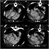
A 7-year-old castrated male Pomeranian dog weighing 5 kg presented with a right-sided continuous murmur without any clinical signs. Thoracic radiographs indicated cardiomegaly and right atrial (RA) bulging. Echocardiography revealed a tunnel originating from the right coronary sinus of Valsalva and terminating in the RA. Contrast echocardiography revealed pulmonary arteriovenous anastomoses. Computed tomography (CT) demonstrated a tortuous shunting vessel that originated from the aorta extending in a ventral direction, ran along the right ventricular wall, and was inserted into the RA. Based on these diagnostic findings, the dog was diagnosed with the aorta-RA tunnel. At the 1-year follow-up visit without treatment, the dog showed no significant change except for mild left ventricular volume overload and mildly decreased contractility. To the best of our knowledge, this is the first case report of an aorta-RA tunnel that has been described in detail using echocardiography and CT in a dog. In conclusion, the aorta-RA tunnel should be included in the clinical differential diagnoses if a right-sided continuous murmur is heard or shunt flow originating from the aortic root is identified.
Keywords: Valsalva; aorta-RA shunt; aorta-RA tunnel; aortic root; canine; cardiovascular anomaly; right atrium; right coronary sinus.
Copyright © 2023 Kim, Ji, Jeong, Lee, Lee and Yoon.
Conflict of interest statement
The authors declare that the research was conducted in the absence of any commercial or financial relationships that could be construed as a potential conflict of interest.
Figures


Similar articles
-
Case report: Echocardiographic and computed tomographic features of congenital bronchoesophageal artery hypertrophy and fistula in a dog.Front Vet Sci. 2024 May 22;11:1400076. doi: 10.3389/fvets.2024.1400076. eCollection 2024. Front Vet Sci. 2024. PMID: 38840636 Free PMC article.
-
A case of aorta-right atrial tunnel presented with an asymptomatic murmur.Korean Circ J. 2013 Sep;43(9):640-3. doi: 10.4070/kcj.2013.43.9.640. Epub 2013 Sep 30. Korean Circ J. 2013. PMID: 24174967 Free PMC article.
-
Aorta-right atrial tunnel.Tex Heart Inst J. 2010;37(4):480-2. Tex Heart Inst J. 2010. PMID: 20844628 Free PMC article.
-
Surgical correction of an aortico-left ventricular tunnel originating from the left aortic sinus with a left coronary artery anomaly.J Card Surg. 2015 Jan;30(1):108-10. doi: 10.1111/jocs.12466. Epub 2014 Nov 14. J Card Surg. 2015. PMID: 25395189 Review.
-
Acute rupture of a sinus of Valsalva aneurysm into the right atrium: a case report and a narrative review.BMC Cardiovasc Disord. 2020 Feb 18;20(1):84. doi: 10.1186/s12872-020-01383-7. BMC Cardiovasc Disord. 2020. PMID: 32070284 Free PMC article. Review.
Cited by
-
Case report of an incidental left coronary artery to main pulmonary artery fistula in a dog.BMC Vet Res. 2025 Aug 4;21(1):503. doi: 10.1186/s12917-025-04730-y. BMC Vet Res. 2025. PMID: 40760024 Free PMC article.
-
Case report: Echocardiographic and computed tomographic features of congenital bronchoesophageal artery hypertrophy and fistula in a dog.Front Vet Sci. 2024 May 22;11:1400076. doi: 10.3389/fvets.2024.1400076. eCollection 2024. Front Vet Sci. 2024. PMID: 38840636 Free PMC article.
References
-
- Iaizzo PA. Handbook of cardiac anatomy, physiology, and devices. Berlin/Heidelberg, Germany: Springer Science & Business Media; (2010).
-
- Garncarz M, Parzeniecka-Jaworska M, Szaluś-Jordanow O. Congenital heart defects in dogs: a retrospective study of 301 dogs. Med Weter. (2017) 73:651–6. doi: 10.21521/mw.5784 - DOI
Publication types
LinkOut - more resources
Full Text Sources
Miscellaneous

