

doi: 10.1021/acsmedchemlett.3c00104.
eCollection 2023 Jul 13.
Discovery of New Binders for DCAF1, an Emerging Ligase Target in the Targeted Protein Degradation Field
Affiliations
- PMID: 37465299
- PMCID: PMC10350940
- DOI: 10.1021/acsmedchemlett.3c00104
Item in Clipboard
Discovery of New Binders for DCAF1, an Emerging Ligase Target in the Targeted Protein Degradation Field
ACS Med Chem Lett.
.
Abstract
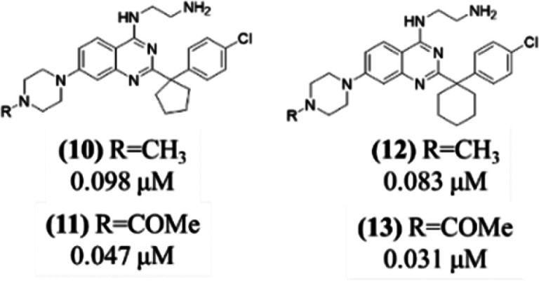
In this study, we describe the rapid identification of potent binders for the WD40 repeat domain (WDR) of DCAF1. This was achieved by two rounds of iterative focused screening of a small set of compounds selected on the basis of internal WDR domain knowledge followed by hit expansion. Subsequent structure-based design led to nanomolar potency binders with a clear exit vector enabling DCAF1-based bifunctional degrader exploration.
© 2023 American Chemical Society.
Conflict of interest statement
The authors declare no competing financial interest.
Figures
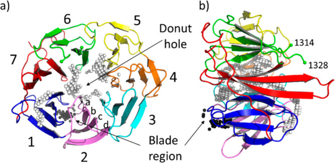
(a) Top view and (b) side view of the crystal structure
(PDB ID: 4CC9) of the human E3
ligase substrate adaptor DCAF1 extracted from the ternary complex
with the viral accessory protein X (Vpx) and the carboxy-terminal
region of human SAMHD1 (not shown). The “top” and “bottom”
surfaces are displayed on the right and left of panel (b), respectively.
Residues 1315–1327 are not visible in the crystal structure.
The Cα atom of the last visible residues, 1314 and 1328 belonging
to blade 6, are marked with a green sphere. The white and black spheres
indicate the two pocket locations as identified by SiteMap and fpocket.
The white spheres correspond to the large ligandable donut hole pocket.
The black spheres indicate the location of the blade region, an additional
less druggable cavity that lies between blade 1 (colored in blue)
and blade 2 (in pink). Each blade (numbered in the picture from 1
to 7) is constituted by a four-stranded antiparallel β-sheet.
Strands are labeled from (a) to (d) from the inside toward the outside
of the propeller.
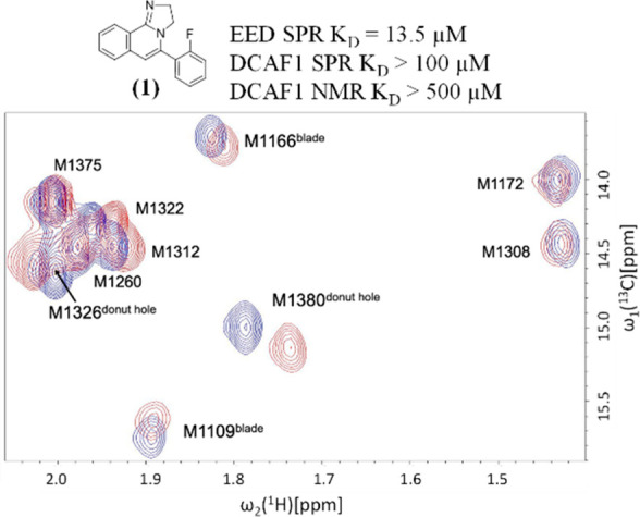
Overlay of the methyl region of 2D [13C,1H]-HMQC
spectra of selectively 13Cε-methionine
labeled DCAF1 in the absence (blue) and in the presence of the compound 1 (red). The concentrations of the protein and the compound
were 15 μM and 480 μM, respectively.
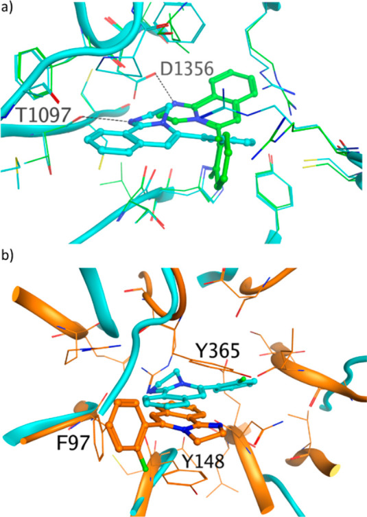
(a) Compound 1 binds to the central donut
cavity of
DCAF1 in two orientations. In both crystal structures, the protonated
imidazoline moiety makes a hydrogen bond, with the carbonyl of T1097
in pose 1 (PDB ID: 8OG6, cyan) and with the carboxyl moiety of D1356
side chain in pose 2 (PDB ID: 8OG5, green). The hydrogen bonds are
shown as dashed black lines. (b) Overlay of crystal structure of 1 bound to DCAF1 (PDB ID: 8OG6, cyan) and to EED (PDB ID:
5H25, orange). The three residues forming the aromatic cages in EED
are labeled.
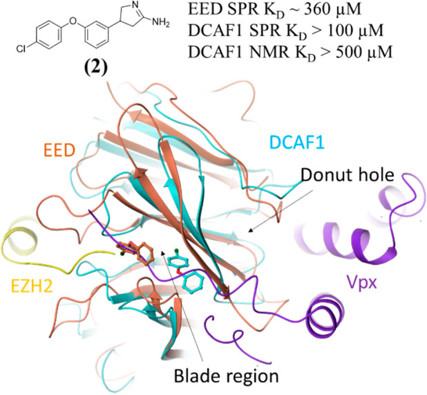
Crystal structures of 2 bound to the blade region
of DCAF1 (PDB ID: 8OG7, cyan) and of EED (orange). Only the most buried
portion of the ligand is built in the density in both crystal structures.
The two crystal structures are overlaid with the DCAF1-Vpx-SAMHD1
crystal structure (PDB ID: 4CC9), from which only the Vpx protein
is shown in violet, and with the EED-1-EZH2 crystal structure
(PDB ID: 5H25), from which only the EZH2 protein is shown in yellow.
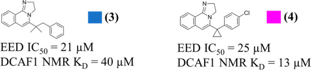
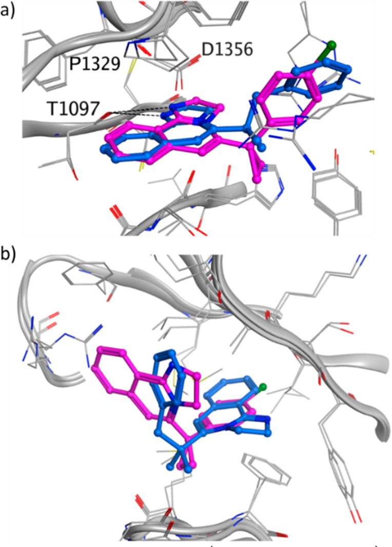
Crystal structure of 3 (PDB ID:
8OG8, blue) and 4 (PDB ID: 8OG9, purple) in (a) the donut
hole cavity and
in (b) the blade region. For 3 only one of the blade
poses is shown. The other pose has the tricyclic core flipped by 180°.
The DCAF1 protein is shown in gray, and the hydrogen bonds are shown
as dashed black lines.
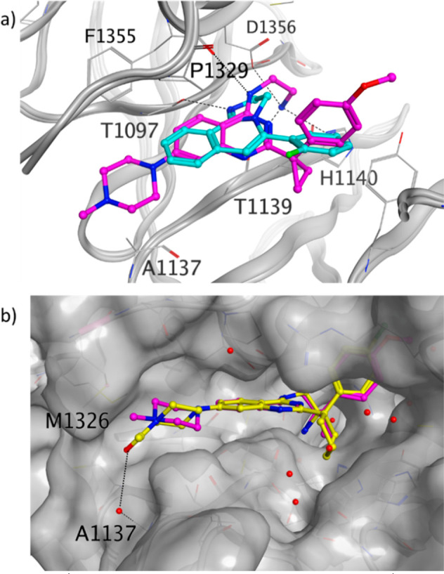
(a) DCAF1 crystal structure
in complex with 8 (PDB
ID: 8OGB, purple) overlaid with the DCAF1 crystal structure in complex
with 1 (PDB ID: 8OG6, cyan). (b) DCAF1 crystal structure
in complex with 11 (PDB ID: 8OGC, yellow) overlaid with
the DCAF1 crystal structure in complex with 8 (purple),
of which only the ligand is shown. The DCAF1 protein surface is shown
in gray, and the hydrogen bonds are shown as dashed black lines.
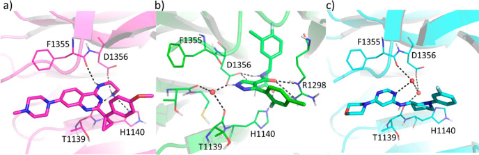
DCAF1
crystal structures in complex with (a) compound 8 (PDB
ID: 8OGB) and (b) compound 26e of reference (18) (PDB ID: 8F8E). (c) DCAF1
crystal structure deposited in PDB under the code 7SSE. Hydrogen bonds
are shown as dashed black lines, and water molecules are shown as
red spheres.
References
-
- Xu C.; Bian C.; Yang W.; Galka M.; Ouyang H.; Chen C.; Qiu W.; Liu H.; Jones A. E.; MacKenzie F.; Pan P.; Li S. S.-C.; Wang H.; Min J. Binding of different histone marks differentially regulates the activity and specificity of polycomb repressive complex 2 (PRC2). Proc. Natl. Acad. Sci. U. S. A. 2010, 107, 19266–19271. 10.1073/pnas.1008937107. - DOI - PMC - PubMed
-
- Tao Y.; Remillard D.; Vinogradova E. V.; Yokoyama M.; Banchenko S.; Schwefel D.; Melillo B.; Schreiber S. L.; Zhang X.; Cravatt B. F. Targeted Protein Degradation by Electrophilic PROTACs that Stereoselectively and Site-Specifically Engage DCAF1. J. Am. Chem. Soc. 2022, 144, 18688–18699. 10.1021/jacs.2c08964. - DOI - PMC - PubMed
-
- Huang Y.; Zhang J.; Yu Z.; Zhang H.; Wang Y.; Lingel A.; Qi W.; Gu J.; Zhao K.; Shultz M. D.; Wang L.; Fu X.; Sun Y.; Zhang Q.; Jiang X.; Zhang J.; Zhang C.; Li L.; Zeng J.; Feng L.; Zhang C.; Liu Y.; Zhang M.; Zhang L.; Zhao M.; Gao Z.; Liu X.; Fang D.; Guo H.; Mi Y.; Gabriel T.; Dillon M. P.; Atadja P.; Oyang C. Discovery of First-in-Class, Potent, and Orally Bioavailable Embryonic Ectoderm Development (EED) Inhibitor with Robust Anticancer Efficacy. J. Med. Chem. 2017, 60, 2215–2226. 10.1021/acs.jmedchem.6b01576. - DOI - PubMed
LinkOut - more resources
Full Text Sources
Other Literature Sources
Chemical Information
Molecular Biology Databases
