Review
. 2023 Jul 12:6:25152564231183898.
doi: 10.1177/25152564231183898. eCollection 2023 Jan-Dec.

The Organization and Function of the Phagophore-ER Membrane Contact Sites

Affiliations
Review

The Organization and Function of the Phagophore-ER Membrane Contact Sites

Prado Vargas Duarte et al. Contact (Thousand Oaks). .

Abstract

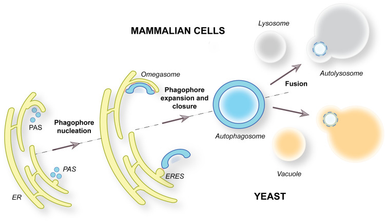
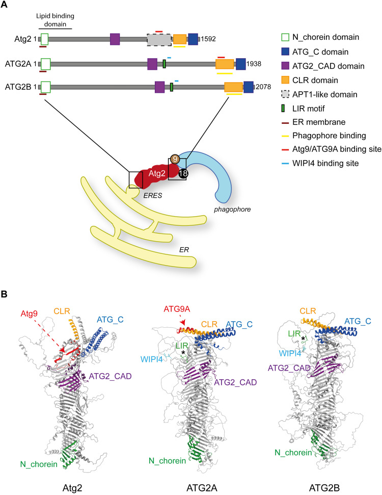
Macroautophagy is characterized by the de novo formation of double-membrane vesicles termed autophagosomes. The precursor structure of autophagosomes is a membrane cistern called phagophore, which elongates through a massive acquisition of lipids until closure. The phagophore establishes membrane-contact sites (MCSs) with the endoplasmic reticulum (ER), where conserved ATG proteins belonging to the ATG9 lipid scramblase, ATG2 lipid transfer and Atg18/WIPI4 β-propeller families concentrate. Several recent in vivo and in vitro studies have uncovered the relevance of these proteins and MCSs in the lipid supply required for autophagosome formation. Although important conceptual advances have been reached, the functional interrelationship between ATG9, ATG2 and Atg18/WIPI4 proteins at the phagophore-ER MCSs and their role in the phagophore expansion are not completely understood. In this review, we describe the current knowledge about the structure, interactions, localizations, and molecular functions of these proteins, with a particular emphasis on the yeast Saccharomyces cerevisiae and mammalian systems.

Keywords: ATG protein; ATG2; ATG9; Atg18/WIPI4; autophagosome; autophagy; lipid transfer; phagophore; scramblase; tethering.


Conflict of interest statement

The author(s) declared no potential conflicts of interest with respect to the research, authorship, and/or publication of this article.

Figures

Figure 1.
Figure 1.
Overview of the Autophagy Pathway. Autophagy is characterized by the de novo formation of double-membrane vesicles called autophagosomes. This process starts with the nucleation of the phagophore, which is probably characterized by the heterotypic fusion of vesicles. The generation of phagophores occurs at the PAS, which is located in close proximity of both the ER and vacuole in yeast (bottom panel), or within omegasomes, a specialized subdomain of the ER, in mammalian cells (upper panel). The phagophores forms MCSs with the ER-exit sites (ERES) in yeast or directly with the ER at the omegasomes in mammals. The phagophore elongation culminates when its extremities fuse together through fission, leading to the formation of an autophagosome. Complete autophagosomes then fuse with vacuoles in yeast and late endosomes/lysosomes in mammalian cells. The autophagosomal cargoes are degraded in the interior of these lytic compartment by resident hydrolases.
Figure 2.
Figure 2.
Structural characteristics of Atg9 and ATG9A. (A) Topological representation of yeast Atg9 and mammalian ATG9A. Atg9 and ATG9A possess four transmembrane domains and two segments partially embedded into the membrane (brown cylinders), which are interconnected by loops (black lines) that differ in amino acid length between the two species. The N- and C-termini of the two proteins, which are coloured in green and red, respectively, are oriented toward the cytosol and they also differ in amino acid length quite remarkably between species. (B) AlphaFold (Jumper et al., 2021; Varadi et al., 2022) prediction of Atg9 and ATG9A protomers. The transmembrane domains of each monomer are coloured in brown, while the cytoplasmic and luminal parts connecting them are in black. N and C termini are coloured as in A. (C) Lateral view of the Atg9/ATG9A trimers. The sequences of the Atg9 and ATG9A were obtained from the UniProt database (https://www.uniprot.org/) and used as template. The modelling of the trimeric complex was performed using the online SWISS-MODEL software (https://swissmodel.expasy.org/interactive) and the resulting structure was coloured using the ChimeraX software (https://www.cgl.ucsf.edu/chimera/) (Pettersen et al., 2004). The structures cover the sequences between amino acids 296 and 783, and between 36 and 352 of Atg9 and ATG9A, respectively. The transmembrane domains of each protomer are highlighted in brown, yellow and orange, while the cytoplasmic domains are in different blue colours. On the right of ATG9A, the interaction between the transmembrane domains of adjacent monomers is highlighted through enlargement. (D) Top view of the homotrimeric structure of Atg9 and ATG9A, obtained by rotating 90 degrees the models presented in Panel C. Lateral pores (LP) are pointed out with dashed red lines, while the central pore (CP) is highlighted in the middle of both structures.
Figure 3.
Figure 3.
Structural Characteristics of Atg2 and its ATG2A/ATG2B Homologs. (A) The domain organization of ATG2 protein consists of a N_Chorein domain located at their N-terminus (green rectangle), in which a Glycerophospholipid binding domain resides. In the middle, Atg2/ATG2 proteins contain a ATG2_CAD domain (purple rectangle), while the CLR region and the ATG_C domain (orange and blue rectangle, respectively) are located at the C-terminus. The membrane binding site responsible of the attachment with the phagophore is within the CLR domain and represented by a yellow line. In contrast to the mammalian ATG2 proteins, Atg2 possesses an APT1-like domain (grey), which is flanking the ATG2_CAD and CLR Motifs. The mammalian ATG2 proteins have one conserved LIR that is represented by a green rectangle in the diagram. As highlighted in the drawing, the N-terminus of the Atg2/ATG2 protein is required for their binding to the ER/ERES while their C-terminus is responsible for the localization at the end of the phagophore and interaction with the Atg9/ATG9A and Atg18/WIPI4 proteins. (B) The structures of Atg2, ATG2A and ATG2B predicted by AlphaFold and coloured based on the diagram in panel A using the ChimeraX Software. The location and name of the different domains is indicated across the structures. The conserved LIR domains in ATG2A and ATG2B are highlighted by asterisks and the amino acids are coloured in green. The Putative WIPI4 binding site in ATG2A and ATG2B is underlined by colouring the amino acids in light blue. The binding region of Atg9 in Atg2 and that of ATG9A in ATG2A are pointed with red dash lines and coloured in the same colour in the structures.
Figure 4.
Figure 4.
Structural Characteristics of Atg18 and WIPI4. (A) The domain organization of Atg18 and WIPI4 is characterized by seven β-propeller blades connected by loops, highlighted with different colours. These proteins function as PtdIns3P effectors and the conserved FRRG/LRRG PtdIns3P-binding motif is indicated. A recent study has shown the presence of the unique 7AB loop (light green) in Atg18. Moreover, the 6CD loop (light blue) is longer in Atg18 than WIPI4. (B) AlphaFold prediction of Atg18 and WIPI4 structures analysed using ChimeraX software are presented. Each blade and loop were coloured based on the domain diagram shown in Panel A. The binding sites for Atg2 and ATG2A/ATG2B are marked by a red arc line in Atg18 and WIPI4, respectively. Similarly, the FRRG and LRRG motif of Atg18 and WIPI4 are highlighted with a black arc line and a dashed arrow, respectively. The 6CD (light blue) 7AB (light green) loops are also indicated.
Figure 5.
Figure 5.
Putative Molecular Function of ATG9, ATG2 and Atg18/WIPI4 Proteins at the Phagophore-ER MCSs. The ER is considered as a key lipids source during phagophore expansion. The current model is that ATG2 proteins localize to the extremities of the phagophore together with ATG9 proteins and Atg18/WIPI4. Atg18/WIPI4 and possibly ATG2 proteins bind to PtdIns3P (orange lipids). ATG2 proteins also bind the ER/ERES through a still unclear mechanism (question mark), and thus they have a key role in establishing the phagophore-ER MCSs. The oval structures on the ER represent the putative ATG2 protein binding partners, which experimental evidence has indicated to be ERES components although which one(s) of them is directly interacting with the ATG2 proteins remains to be determined. Although it remains unknown what is providing the force for the flow of lipids, those are transferred from the ER to the phagophore by ATG2 proteins (black arrow), a process that also requires the presence of Atg18/WIPI4 at the phagophore-ER MCSs. Lipids from ATG2 proteins may or may not be delivered to ATG9 scramblases, which translocate bidirectionally the lipids across the phagophore membrane bilayer, maintaining the symmetric distribution of lipids and ultimately allowing phagophore expansion.

References

    1. Bakula D, Muller AJ, Zuleger T, Takacs Z, Franz-Wachtel M, Thost AK, Brigger D, Tschan MP, Frickey T, Robenek H, … (2017). WIPI3 And WIPI4 beta-propellers are scaffolds for LKB1-AMPK-TSC signalling circuits in the control of autophagy. Nature Communications 8, 15637. doi: 10.1038/ncomms15637 - DOI - PMC - PubMed
    1. Barth H, Meiling-Wesse K, Epple UD, Thumm M. (2001). Autophagy and the cytoplasm to vacuole targeting pathway both require Aut10p. FEBS Letters 508, 23–28. doi: 10.1016/s0014-5793(01)03016-2 - DOI - PubMed
    1. Baskaran S, Ragusa MJ, Boura E, Hurley JH. (2012a). Two-site recognition of phosphatidylinositol 3-phosphate by PROPPINs in autophagy. Molecular Cell 47, 339–348. doi: 10.1016/j.molcel.2012.05.027. - DOI - PMC - PubMed
    1. Baskaran S, Ragusa MJ, Hurley JH. (2012b). How Atg18 and the WIPIs sense phosphatidylinositol 3-phosphate. Autophagy 8, 1851–1852. doi: 10.4161/auto.22077. - DOI - PMC - PubMed
    1. Behrends C, Sowa ME, Gygi SP, Harper JW. (2010). Network organization of the human autophagy system. Nature 466, 68–76. doi: 10.1038/nature09204. - DOI - PMC - PubMed

LinkOut - more resources