. 2022 May;37(3):220-230.
doi: 10.1177/08839115221095154. Epub 2022 May 17.

Galloylated proanthocyanidins in dentin matrix exhibit biocompatibility and induce differentiation in dental stem cells

Affiliations

Galloylated proanthocyanidins in dentin matrix exhibit biocompatibility and induce differentiation in dental stem cells

Daniel Kulakowski et al. J Bioact Compat Polym. 2022 May.

Abstract

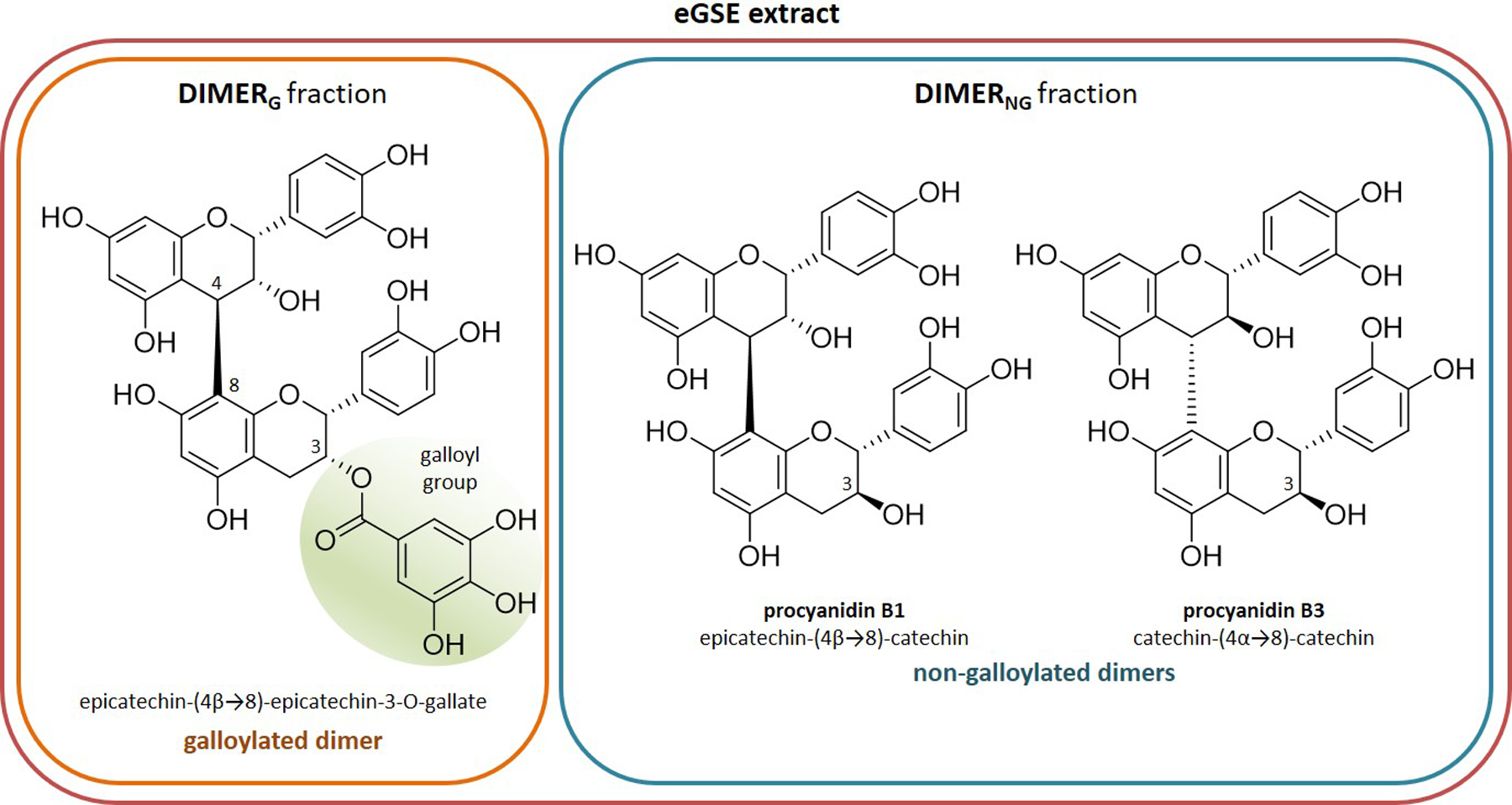
Aim: Grape seed extract contains a complex mixture of proanthocyanidins (PACs), a plant biopolymer used as a biomaterial to improve reparative and preventive dental therapies. Co-polymerization of PACs with type I collagen mechanically reinforces the dentin extracellular matrix. This study assessed the biocompatibility of PACs from grape seed extract on dental pulp stem cells (DPSCs) in a model simulating leaching through dentin to the pulp cavity. The aim was to determine the type of PACs (galloylated vs. non-galloylated) within grape seed extract that are most compatible with dental pulp tissue.

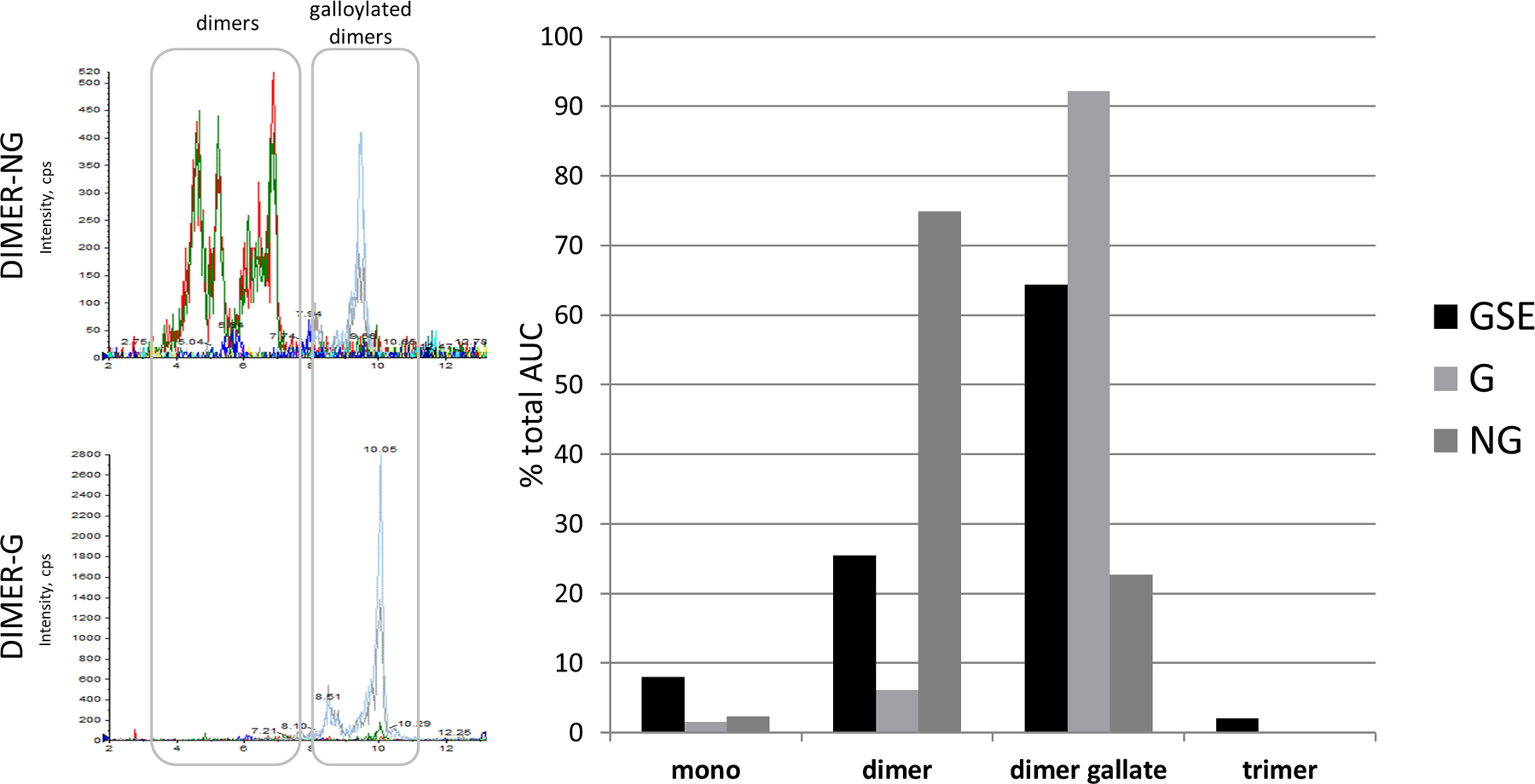
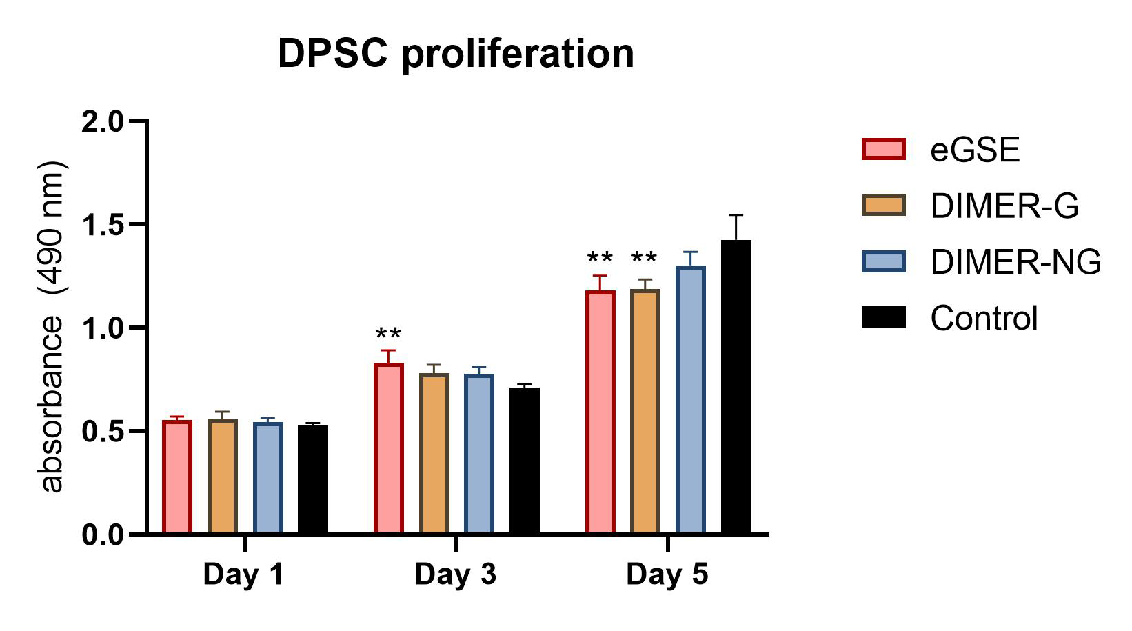
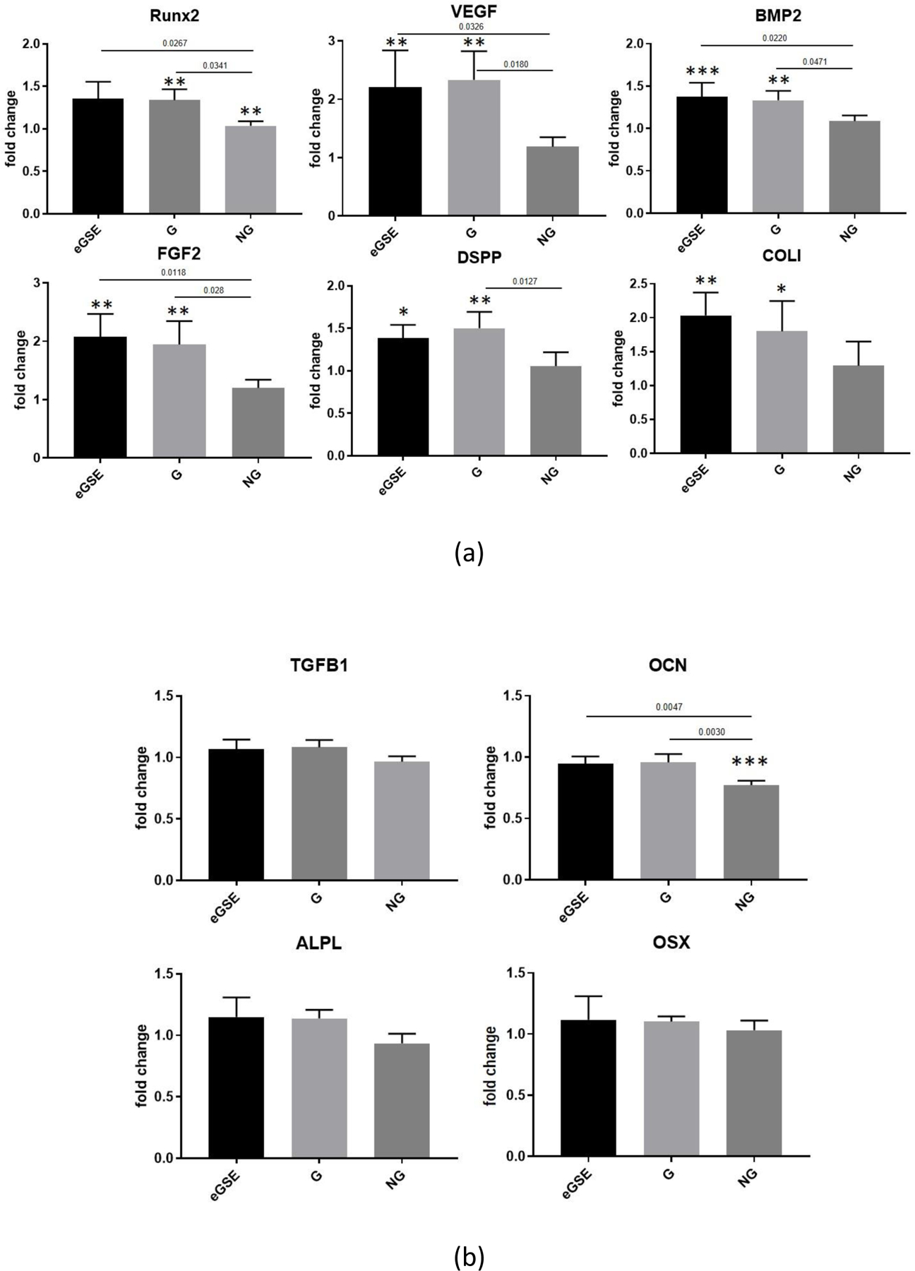
Methodology: Human demineralized dentin was treated with selectively-enriched dimeric PACs prepared from grape seed extract using liquid-liquid chromatography. DPSCs were cultured within a 2D matrix and exposed to PAC-treated dentin extracellular matrix. Cell proliferation was measured using the MTS assay and expression of odontoblastic genes was analyzed by qRT-PCR. Categorization of PACs leaching from dentin was performed using HPLC-MS.

Results: Enriched dimeric fractions containing galloylated PACs increased the expression of certain odontoblastic genes in DPSCs, including Runt-related transcription factor 2 (RUNX2), vascular endothelial growth factor (VEGF), bone morphogenetic protein 2 (BMP2), basic fibroblast growth factor (FGF2), dentin sialophosphoprotein (DSPP) and collagen, type I, alpha 1 (COLI). Galloylated dimeric PACs also exhibited minor effects on DPSC proliferation, resulting in a decrease compared to control after five days of treatment. The non-galloylated dimer fraction had no effect on these genes or on DPSC proliferation.

Conclusions: Galloylated PACs are biocompatible with DPSCs and may exert a beneficial effect on cells within dental pulp tissue. The observed increase in odontoblastic genes induced by galloylated PACs together with a decrease in DPSC proliferation is suggestive of a shift toward cell differentiation. This data supports the use of dimeric PACs as a safe biomaterial, with galloylated dimeric PACs exhibiting potential benefits to odontoblasts supporting dentin regeneration.

Keywords: biocompatibility; cell differentiation; cell viability; dental pulp stem cells; dentin; plant biopolymers; proanthocyanidins.


Conflict of interest statement

Declaration of Conflicting Interests The Authors declare that there is no conflict of interest.

Figures

Figure 1.
Figure 1.
Structure of representative, major, components of eGSE extract and DIMERG and DIMERNG fractions. Galloyl group is highlighted. Epicatechin-(4β → 8)-epicatechin-3-O-gallate is the major component of DIMERG and procyanidin B1 and procyanidin B3 are the major components of DIMERNG [4].
Figure 2.
Figure 2.
HPLC-MS analysis of PACs leaching from treated dentin beams into culture media. Media was withdrawn after 5 days of exposure to treated beams. (Left Panel) Extracted ion chromatograms of dimers (m/z- 577.20 → 407.10) and galloylated dimers (m/z- 729.20 → 577.20, 729.20 → 407.10). (Right Panel) Bar graph representing % contribution of each size PAC to the total AUC (area under the curve) of MRM (multiple-reaction monitoring) chromatograms, respective of treatment. Mono = monomer.
Figure 3.
Figure 3.
Proliferation of DPSCs exposed to dentin treated with eGSE and refined extracts in model of dentin-pulp complex after 1, 3 and 5 days. n = 4 culture replicates per treatment. Error bars represent standard deviation. **, p < 0.01 compared to control, by Tukey’s test after 1-way ANOVA.
Figure 4.
Figure 4.
Cell viability of DPSCs exposed to eGSE at 100, 10 and 1 ug/mL direct treatment after 24 hrs. Live cells stained green by calcein-AM. Dead cells stained red by ethidium homodimer. Shown at 20× magnification.
Figure 5
Figure 5
(a) Expression of odontoblastic genes after DPSCs were exposed to eGSE, DIMERG, and DIMERNG fractions for 5days. Displayed as relative gene expression compared to negative control (fold change). Genes upregulated by leaching of PACs include RUNX2 (Runt-related transcription factor 2), VEGF (vascular endothelial growth factor), BMP2 (bone morphogenetic protein 2), FGF2 (basic fibroblast growth factor), DSPP (dentin sialophosphoprotein) and COLI (collagen, type I, alpha 1). eGSE = enriched grape seed extract, G = DIMERG, NG = DIMERNG, p-values displayed are between treatments, using fold change values. Error bars represent standard deviation. *p<0.05; **p<0.01, ***p<.001 compared to control, using dCq values. Comparisons made by Tukey’s test after 1-way ANOVA. n=4 culture replicates per treatment. (b) Expression of odontoblastic genes after DPSCs were exposed to GSE, DIMERG and DIMERNG fractions for 5days. Displayed as relative gene expression compared to negative control. Genes downregulated or not changed by leaching of PACs include TGF-β1 (transforming growth factor beta 1), ALPL (alkaline phosphatase), OSX (osteoblast transcription factor Sp7) and OCN (osteocalcin). eGSE = enriched grape seed extract, G = DIMERG, NG = DIMERNG. p-values displayed are between treatments, using fold change values. Error bars represent standard deviation. ***p<.001 compared to control, using dCq values. Comparisons made by Tukey’s test after 1-way ANOVA. n=4 culture replicates per treatment.

Similar articles

Cited by

References

    1. Leme-Kraus AA, Aydin B, Vidal CMP, et al. Biostability of the Proanthocyanidins-Dentin Complex and Adhesion Studies. J Dent Res 2017; 96: 406–412. - PMC - PubMed
    1. Aguiar TR, Vidal CMP, Phansalkar RS, et al. Dentin biomodification potential depends on polyphenol source. J Dent Res 2014; 93: 417–422. - PMC - PubMed
    1. Vidal CMP, Aguiar TR, Phansalkar R, et al. Galloyl moieties enhance the dentin biomodification potential of plant-derived catechins. Acta Biomater 2014; 10: 3288–3294. - PMC - PubMed
    1. Phansalkar RS, Nam J-W, Chen S-N, et al. A galloylated dimeric proanthocyanidin from grape seed exhibits dentin biomodification potential. Fitoterapia 2015; 101: 169–178. - PMC - PubMed
    1. Geurtsen W. Biocompatibility of resin-modified filling materials. Crit Rev Oral Biol Med 2000; 11: 333–355. - PubMed

LinkOut - more resources