

Rare manifestation of familial vitreous amyloidosis caused by Gly103Arg transthyretin
- PMID: 37465499
- PMCID: PMC10333243
- DOI: 10.18240/ijo.2023.07.14
Rare manifestation of familial vitreous amyloidosis caused by Gly103Arg transthyretin
Abstract
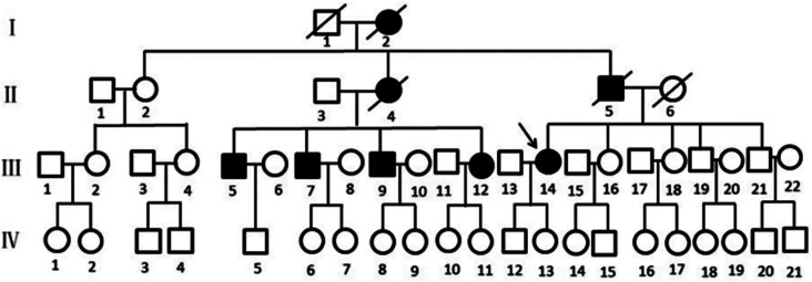
Aim: To identify and analyze the genotype of the patients with special ocular manifestations of familial vitreous amyloidosis (FVA) in a Chinese Han family.
Methods: Pars plana vitrectomy (PPV) surgery was performed on a 52-year-old Chinese woman presented with vitreous amyloidosis and progressive visual impairment, without evidence of cardiac, renal, gastrointestinal, central nervous system or peripheral nervous system dysfunction. During the surgery, the patient presented with a gray-white dense and thick cotton wool-like change in the vitreous body, accompanied by complete retinal detachment. Additionally, hard, free and movable yellow-white deposits were observed in the posterior pole and surrounding retina, the vitreous and subretinal deposits were examined by Congo red staining and immunohistochemical pathological examination, and whole exome sequencing was performed on blood samples from the patient and her cousin.
Results: During the operation, it was discovered that there was a complete detachment of the retina and a significant amount of hard, free-floating yellow-white deposits were observed beneath the posterior pole and surrounding retina. This is an exceedingly rare ocular manifestation. Pathological examination of the vitreous and subretinal deposit specimens revealed positive Congo red staining, as well as elevated vascular endothelial growth factor (VEGF) expression in vascular endothelial cells within the sediment specimens upon immunohistochemical examination. The patient and her cousin both exhibited a heterozygous mutation in Glyl03Arg within the transthyretin (TTR) gene, resulting in a substitution of glycine (Gly) at position 103 with arginine (Arg).
Conclusion: FVA may present with various ocular manifestations, but panretinal detachment is a rare occurrence. In cases where retinal detachment persists for an extended period of time, amyloid deposits may form under the retina through retinal tears, leading to subretinal deposits that can impede retinal reattachment and negatively impact visual prognosis. Elevated levels of VEGF in the eyes of FVA patients may indicate an overexpression state, necessitating careful postoperative follow-up. The heterozygous mutation Gly103Arg may represent a unique pathogenic site in Chinese individuals.
Keywords: Gly103Arg; familial vitreous amyloidosis; transthyretin gene; vascular endothelial growth factor.
International Journal of Ophthalmology Press.
Figures


Similar articles
-
Transthyretin Ala36Pro mutation in a Chinese pedigree of familial transthyretin amyloidosis with elevated vitreous and serum vascular endothelial growth factor.Exp Eye Res. 2013 May;110:44-9. doi: 10.1016/j.exer.2013.02.005. Epub 2013 Feb 21. Exp Eye Res. 2013. PMID: 23438977
-
Case Report: Hereditary transthyretin (ATTRv) amyloidosis: The p.G103R mutation of the transthyretin gene in a Han Chinese family is associated with vitreous hemorrhage.Front Genet. 2022 Sep 15;13:972501. doi: 10.3389/fgene.2022.972501. eCollection 2022. Front Genet. 2022. PMID: 36186469 Free PMC article.
-
Vitreous Amyloidosis: Ocular, Systemic, and Genetic Insights.Ophthalmology. 2017 Jul;124(7):1014-1022. doi: 10.1016/j.ophtha.2017.03.011. Epub 2017 Apr 12. Ophthalmology. 2017. PMID: 28412068
-
Pars plana vitrectomy versus scleral buckling for repairing simple rhegmatogenous retinal detachments.Cochrane Database Syst Rev. 2019 Mar 8;3(3):CD009562. doi: 10.1002/14651858.CD009562.pub2. Cochrane Database Syst Rev. 2019. PMID: 30848830 Free PMC article.
-
[The ocular involvement in the transthyretin-related familial amyloid polyneuropathy].Zhonghua Yan Ke Za Zhi. 2017 Oct 11;53(10):783-785. doi: 10.3760/cma.j.issn.0412-4081.2017.10.014. Zhonghua Yan Ke Za Zhi. 2017. PMID: 29050191 Review. Chinese.
Cited by
-
Global research trends in vitreous floaters from 1999 to 2023: a bibliometric analysis.Int Ophthalmol. 2025 Apr 10;45(1):152. doi: 10.1007/s10792-025-03492-8. Int Ophthalmol. 2025. PMID: 40208401
-
Hereditary vitreoretinal amyloidosis with transthyretin Gly83Arg variant, a long-term study.Eye (Lond). 2025 Feb;39(2):345-353. doi: 10.1038/s41433-024-03445-y. Epub 2024 Oct 31. Eye (Lond). 2025. PMID: 39478196
-
The Gly103Arg variant in hereditary transthyretin amyloidosis.Front Neurol. 2024 Sep 9;15:1471131. doi: 10.3389/fneur.2024.1471131. eCollection 2024. Front Neurol. 2024. PMID: 39314866 Free PMC article.
References
-
- Choi KJ, Son KY, Kang SW, et al. Ocular manifestations of asp38ala and thr59lys familial transthyretin amyloidosis. Retina. 2022;42(2):396–403. - PubMed
-
- Rowczenio DM, Noor I, Gillmore JD, et al. Online registry for mutations in hereditary amyloidosis including nomenclature recommendations. Hum Mutat. 2014;35(9):E2403–E2412. - PubMed
-
- Dammacco R, Merlini G, Lisch W, Kivelä TT, Giancipoli E, Vacca A, Dammacco F. Amyloidosis and ocular involvement: an overview. Semin Ophthalmol. 2020;35(1):7–26. - PubMed
LinkOut - more resources
Full Text Sources
Research Materials
Miscellaneous