

Abnormal L5-S1 Facet Joint Orientation as a Harbinger of Degenerative Spondylolisthesis: A Case Report
- PMID: 37465811
- PMCID: PMC10351618
- DOI: 10.7759/cureus.40569
Abnormal L5-S1 Facet Joint Orientation as a Harbinger of Degenerative Spondylolisthesis: A Case Report
Abstract
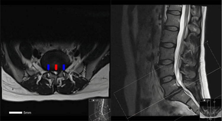
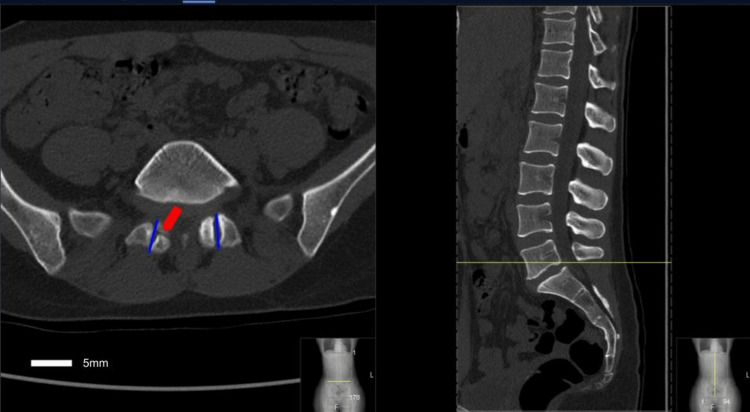
Degenerative spondylolisthesis is a common cause of low back pain and resultant disability in the adult population. The causes of degenerative spondylolisthesis are not entirely understood, though a combination of anatomic and lifestyle factors likely contributes to the development of this pathology. Here, we report a case of a 38-year-old female presenting with low back pain and right lower extremity radiculopathy, found to have degenerative L5-S1 spondylolisthesis, which we postulate developed in part due to the sagittal orientation of her L5-S1 facet joints bilaterally.
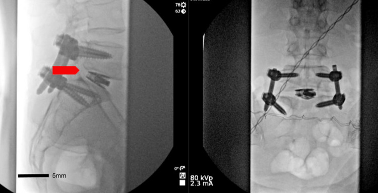
Keywords: injury biomechanics; lumbar spine; neurosurgery; orthopedic surgery; spine surgery; spondylolisthesis; surgery; transforaminal lumbar interbody fusion.
Copyright © 2023, Labak et al.
Conflict of interest statement
The authors have declared that no competing interests exist.
Figures

Similar articles
-
[Adjacent segment degeneration after lumbosacral fusion in spondylolisthesis: a retrospective radiological and clinical analysis].Acta Chir Orthop Traumatol Cech. 2010 Apr;77(2):124-30. Acta Chir Orthop Traumatol Cech. 2010. PMID: 20447355 Czech.
-
Orientation and osteoarthritis of the lumbar facet joint.Clin Orthop Relat Res. 2001 Apr;(385):88-94. doi: 10.1097/00003086-200104000-00015. Clin Orthop Relat Res. 2001. PMID: 11302332
-
Sagittal spinopelvic alignment and body mass index in patients with degenerative spondylolisthesis.Eur Spine J. 2011 May;20(5):713-9. doi: 10.1007/s00586-010-1640-2. Epub 2010 Dec 1. Eur Spine J. 2011. PMID: 21116661 Free PMC article.
-
Is Stand-Alone Anterior Lumbar Interbody Fusion a Safe and Efficacious Treatment for Isthmic Spondylolisthesis of L5-S1?Global Spine J. 2017 Sep;7(6):587-595. doi: 10.1177/2192568217699210. Epub 2017 Jun 1. Global Spine J. 2017. PMID: 28894689 Free PMC article. Review.
-
Acute progression of spondylolysis to isthmic spondylolisthesis in an adult.Spine (Phila Pa 1976). 2002 Aug 15;27(16):E370-2. doi: 10.1097/00007632-200208150-00023. Spine (Phila Pa 1976). 2002. PMID: 12195078 Review.
References
-
- Natural history of degenerative spondylolisthesis. A systematic review and meta-analysis. (Article in press) Atalay B, Gadjradj PS, Sommer FS, Wright D, Rawanduzy C, Ghogawala Z, Härtl R. World Neurosurg. 2023 - PubMed
-
- Pathomechanisms and predisposing factors for degenerative lumbar spondylolisthesis: a narrative review. Yoshihara H. JBJS Rev. 2020;8:0. - PubMed
-
- Degenerative lumbar spondylolisthesis: definition, natural history, conservative management, and surgical treatment. Bydon M, Alvi MA, Goyal A. Neurosurg Clin N Am. 2019;30:299–304. - PubMed
-
- Neurogenic claudication secondary to degenerative spondylolisthesis: is fusion always necessary? Kitchen WJ, Mohamed M, Bhojak M, Wilby M. Br J Neurosurg. 2016;30:662–665. - PubMed
-
- Two-year comprehensive medical management of degenerative lumbar spine disease (lumbar spondylolisthesis, stenosis, or disc herniation): a value analysis of cost, pain, disability, and quality of life: clinical article. Parker SL, Godil SS, Mendenhall SK, Zuckerman SL, Shau DN, McGirt MJ. J Neurosurg Spine. 2014;21:143–149. - PubMed
Publication types
LinkOut - more resources
Full Text Sources