

A case of epiglottic entrapment in a cat
- PMID: 37465985
- PMCID: PMC10508545
- DOI: 10.1002/vms3.1211
A case of epiglottic entrapment in a cat
Abstract
Objective: The objective of this study was to describe a case of epiglottic entrapment in a cat.
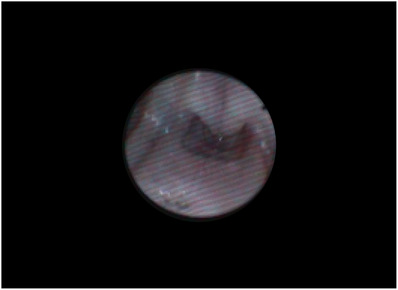
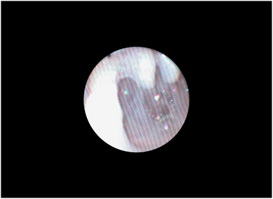
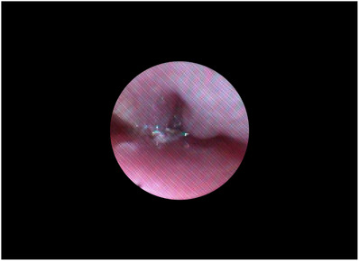
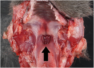
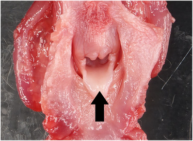
Case summary: A 5-month-old male neutered Russian Blue cat was evaluated for progressive stertorous upper airway sounds, acute onset vestibulopathy and abnormal laryngeal anatomy. Endotracheal intubation was only able to be achieved using videoscopic guidance and identified concern for severe nasopharyngeal stenosis. A computerized tomography scan revealed otitis interna, narrowed nasopharynx and no definitive cause for the stertorous breathing. The cat recovered very slowly from anaesthesia due to concern for airway obstruction following extubation. It was discharged the following day and then passed away at home 2 weeks later. Necropsy revealed that the epiglottis was obscured by 2 cm of redundant mucosal tissue extending from the base of the tongue to the larynx resulting in epiglottic entrapment. Also noted was chronic, severe otitis interna and externa. Upper airway obstruction is suspected to be the cause of sudden death.
New or unique information: To the authors' knowledge, this is the first report of these oropharyngeal anatomic abnormalities in a cat.
Keywords: cartilage; disease; epiglottic; feline; nasopharyngeal.
© 2023 The Authors. Veterinary Medicine and Science published by John Wiley & Sons Ltd.
Conflict of interest statement
The authors declare no conflict of interest.
Figures

References
-
- Ambrosio, A. , & Brigger, M. T. (2012). Pediatric supraglottoplasty. Advances in Oto‐Rhino‐Laryngology, 73, 101–104. - PubMed
-
- Bedford, P. G. (1983). Displacement of the glosso‐epiglottic mucosa in canine asphyxiate disease. Journal of Small Animal Practice, 24(4), 199–207.
-
- Bedwell, J. , & Zalzal, G. (2016). Laryngomalacia. Seminars in Pediatric Surgery, 25(3), 119–122. - PubMed
-
- Boles, C. L. , Raker, C. W. , & Wheat, J. F. (1978). Epiglottic entrapment by arytenoepiglottic folds in the horse. Journal of the American Veterinary Medical Association, 172(3), 338–342. - PubMed
-
- Flanders, J. A. , & Thompson, M. S. (2009). Dyspnea caused by epiglottic retroversion in two dogs. Journal of the American Veterinary Medical Association, 235(11), 1330–1335. - PubMed
Publication types
MeSH terms
LinkOut - more resources
Full Text Sources
Medical
Miscellaneous
