Tumor budding and tumor-infiltrating lymphocytes can predict prognosis in pT1b esophageal squamous cell carcinoma
- PMID: 37466146
- PMCID: PMC10481137
- DOI: 10.1111/1759-7714.15043
Tumor budding and tumor-infiltrating lymphocytes can predict prognosis in pT1b esophageal squamous cell carcinoma
Abstract
Background: Tumor budding (TB) and tumor-infiltrating lymphocyte (TIL) are significant predictive indicators of lymph node metastasis (LNM) and unfavorable prognosis in various tumors. Currently, there is no gold standard for TB and TIL evaluation in esophageal squamous cell carcinoma (ESCC). This study aimed to identify the standard of TB and TIL evaluations and build a predictive model for prognosis among patients with pT1b ESCC.
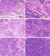
Methods: We retrospectively analyzed the prognostic values of TB and TIL in 150 pT1b ESCC cases. Hematoxylin and eosin (H&E) and immunohistochemistry (IHC) of anti-pan cytokeratin (AE1/AE3) were used to analyze the threshold of TB, and intratumoral TIL and peritumoral TIL (pTIL) were evaluated using the receiver operating characteristic curves (ROC).
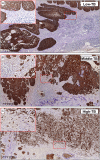
Results: We found that TB in a three-tiered grading system (low-TB: 0-4; middle-TB: 5-15; high-TB: ≥16) displayed an excellent prognosis prediction for LNM and survival based on IHC staining using a 20× objective lens. Low pTIL level (≤20%) was a significant indicator of LNM and unfavorable prognosis (p < 0.05). Moreover, lower tumor location and lymphovascular invasion (LVI) were correlated with an unfavorable prognosis (p < 0.05). A nomogram developed based on TB, pTIL, LVI, and tumor location showed good discrimination, as shown by the area under the ROC and calibration curves.
Conclusion: We therefore recommend identifying TB using a 20× objective lens under IHC staining and TIL adjacent to the tumor. Additionally, a nomogram was built for facilitating individualized prediction of survival for patients with pT1b ESCC.
Keywords: lymph node metastasis; nomogram; prognosis; tumor budding; tumor infiltrating lymphocyte.
© 2023 The Authors. Thoracic Cancer published by China Lung Oncology Group and John Wiley & Sons Australia, Ltd.
Conflict of interest statement
The authors confirm there are no conflicts of interest.
Figures


Similar articles
-
Prognostic significance of lymphovascular invasion in patients with pT1b esophageal squamous cell carcinoma.BMC Cancer. 2023 Apr 22;23(1):370. doi: 10.1186/s12885-023-10858-7. BMC Cancer. 2023. PMID: 37087442 Free PMC article.
-
Immunohistochemical analysis of tumor budding as predictor of lymph node metastasis from superficial esophageal squamous cell carcinoma.Esophagus. 2020 Apr;17(2):168-174. doi: 10.1007/s10388-019-00698-5. Epub 2019 Oct 8. Esophagus. 2020. PMID: 31595396
-
Nomogram for prediction of lymph node metastasis in patients with superficial esophageal squamous cell carcinoma.J Gastroenterol Hepatol. 2020 Jun;35(6):1009-1015. doi: 10.1111/jgh.14915. Epub 2019 Dec 15. J Gastroenterol Hepatol. 2020. PMID: 31674067
-
Risk factors for lymph node metastasis in T1 esophageal squamous cell carcinoma: A systematic review and meta-analysis.World J Gastroenterol. 2021 Feb 28;27(8):737-750. doi: 10.3748/wjg.v27.i8.737. World J Gastroenterol. 2021. PMID: 33716451 Free PMC article.
-
Tumor budding is a prognostic marker for overall survival and not for lymph node metastasis in Oral Squamous Cell Carcinoma - Systematic Review Update and Meta-Analysis.J Oral Biol Craniofac Res. 2024 Jul-Aug;14(4):362-369. doi: 10.1016/j.jobcr.2024.04.013. Epub 2024 May 23. J Oral Biol Craniofac Res. 2024. PMID: 38832296 Free PMC article. Review.
Cited by
-
Risk factors for lymph node metastasis in superficial esophageal squamous cell carcinoma.World J Gastroenterol. 2024 Apr 7;30(13):1810-1814. doi: 10.3748/wjg.v30.i13.1810. World J Gastroenterol. 2024. PMID: 38659479 Free PMC article.
-
Unveiling Another Dimension: Advanced Visualization of Cancer Invasion and Metastasis via Micro-CT Imaging.Cancers (Basel). 2025 Mar 28;17(7):1139. doi: 10.3390/cancers17071139. Cancers (Basel). 2025. PMID: 40227647 Free PMC article.
-
Tumor Budding in Upper Gastrointestinal Carcinomas: A Systematic Review and Meta-Analysis.Cureus. 2024 Sep 29;16(9):e70422. doi: 10.7759/cureus.70422. eCollection 2024 Sep. Cureus. 2024. PMID: 39473660 Free PMC article. Review.
-
Nomograms and prognosis for superficial esophageal squamous cell carcinoma.World J Gastroenterol. 2024 Mar 14;30(10):1291-1294. doi: 10.3748/wjg.v30.i10.1291. World J Gastroenterol. 2024. PMID: 38596490 Free PMC article.
-
Risk factors and a predictive nomogram for lymph node metastasis in superficial esophageal squamous cell carcinoma.World J Gastroenterol. 2023 Dec 21;29(47):6138-6147. doi: 10.3748/wjg.v29.i47.6138. World J Gastroenterol. 2023. PMID: 38186680 Free PMC article.
References
-
- Sung H, Ferlay J, Siegel RL, Laversanne M, Soerjomataram I, Jemal A, et al. Global cancer statistics 2020: GLOBOCAN estimates of incidence and mortality worldwide for 36 cancers in 185 countries. CA Cancer J Clin. 2021;71(3):209–249. - PubMed
-
- Xue L, Ren L, Zou S, Shan L, Liu X, Xie Y, et al. Parameters predicting lymph node metastasis in patients with superficial esophageal squamous cell carcinoma. Mod Pathol. 2015;28(1):161. - PubMed
-
- Kadota K, Nitadori J, Woo KM, Sima CS, Finley DJ, Rusch VW, et al. Comprehensive pathological analyses in lung squamous cell carcinoma: single cell invasion, nuclear diameter, and tumor budding are independent prognostic factors for worse outcomes. J Thorac Oncol. 2014;9(8):1126–1139. - PMC - PubMed
Publication types
MeSH terms
LinkOut - more resources
Full Text Sources
Medical
Miscellaneous

