

KCa 2.2 (KCNN2): A physiologically and therapeutically important potassium channel
- PMID: 37466411
- PMCID: PMC10932612
- DOI: 10.1002/jnr.25233
KCa 2.2 (KCNN2): A physiologically and therapeutically important potassium channel
Abstract
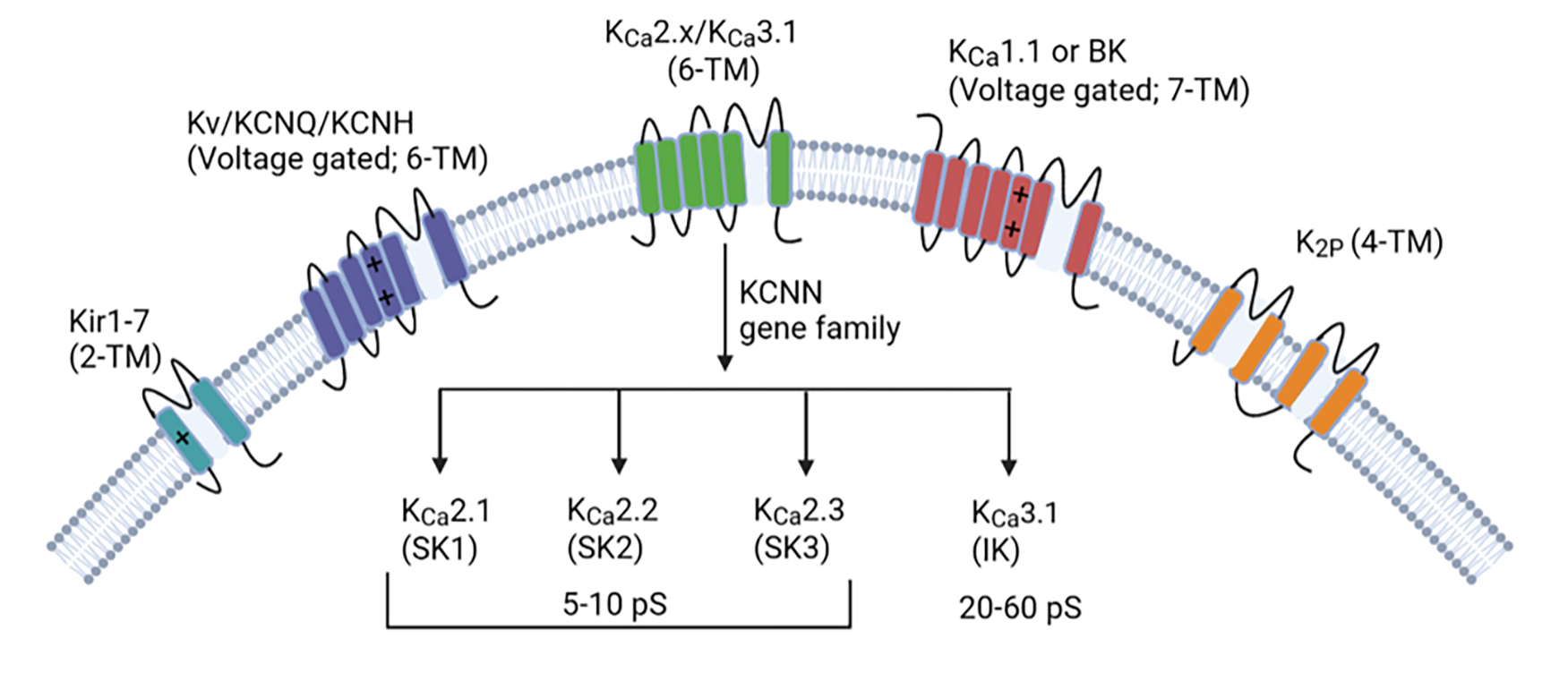
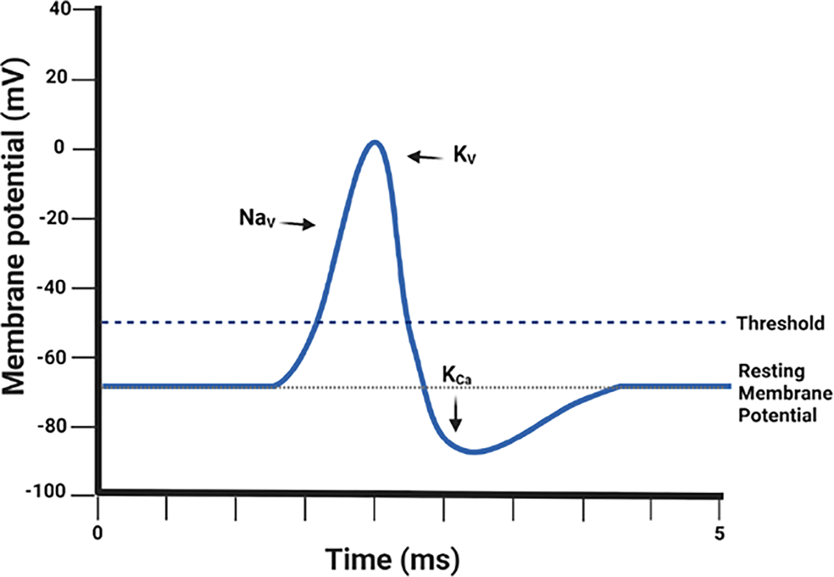
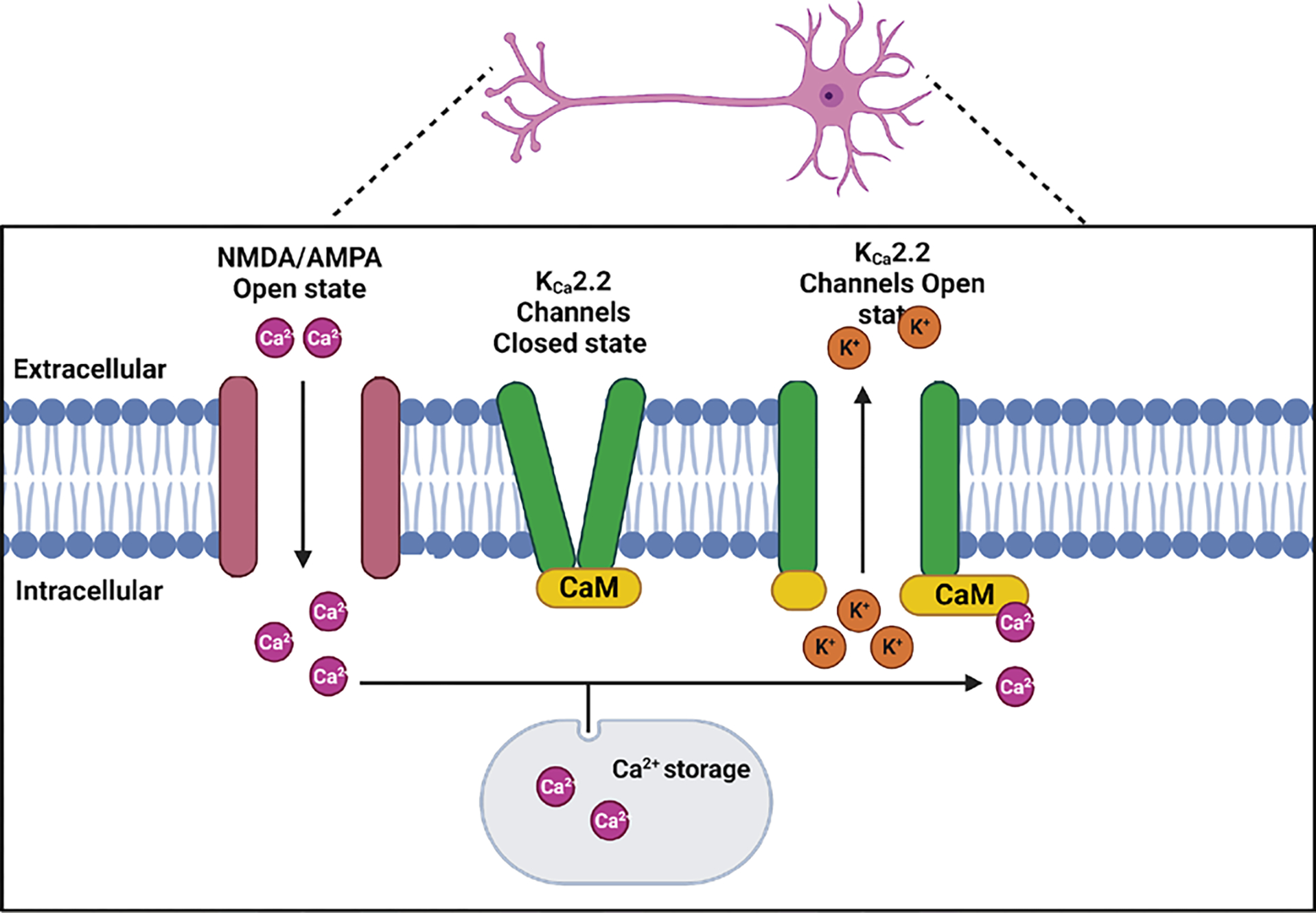
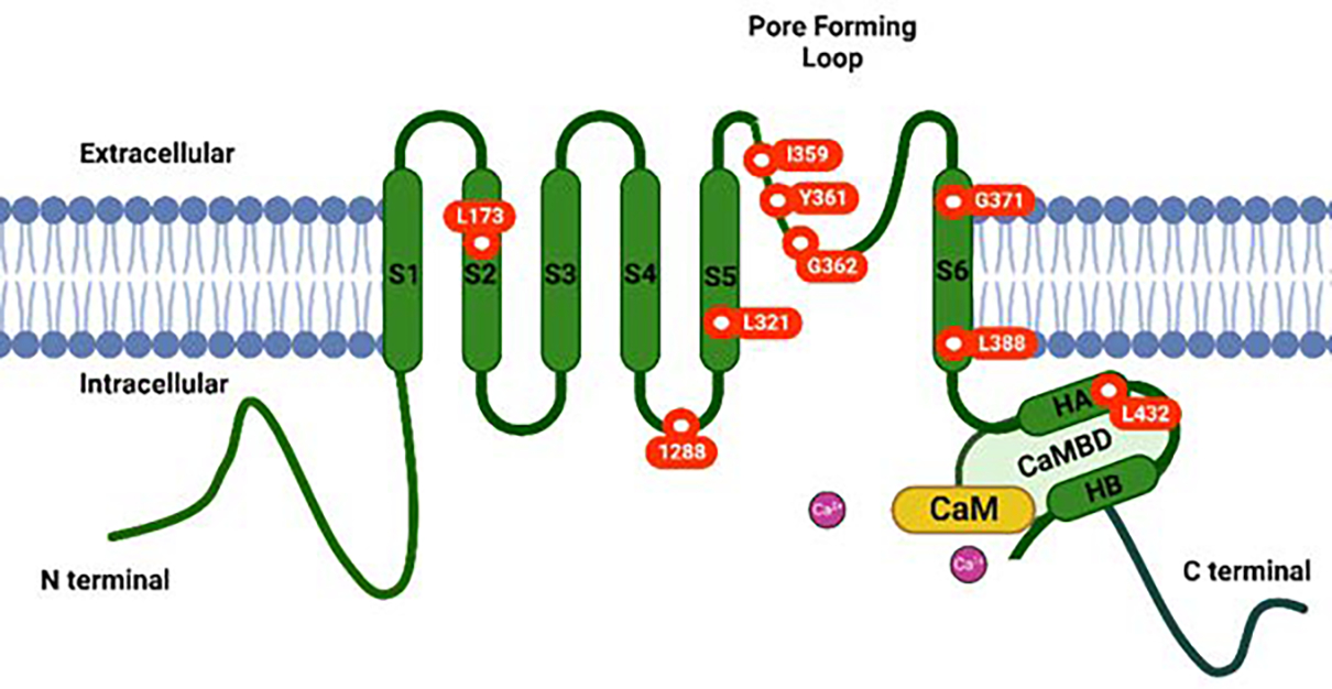
One group of the K+ ion channels, the small-conductance Ca2+ -activated potassium channels (KCa 2.x, also known as SK channels family), is widely expressed in neurons as well as the heart, endothelial cells, etc. They are named small-conductance Ca2+ -activated potassium channels (SK channels) due to their comparatively low single-channel conductance of about ~10 pS. These channels are insensitive to changes in membrane potential and are activated solely by rises in the intracellular Ca2+ . According to the phylogenic research done on the KCa 2.x channels family, there are three channels' subtypes: KCa 2.1, KCa 2.2, and KCa 2.3, which are encoded by KCNN1, KCNN2, and KCNN3 genes, respectively. The KCa 2.x channels regulate neuronal excitability and responsiveness to synaptic input patterns. KCa 2.x channels inhibit excitatory postsynaptic potentials (EPSPs) in neuronal dendrites and contribute to the medium afterhyperpolarization (mAHP) that follows the action potential bursts. Multiple brain regions, including the hippocampus, express the KCa 2.2 channel encoded by the KCNN2 gene on chromosome 5. Of particular interest, rat cerebellar Purkinje cells express KCa 2.2 channels, which are crucial for various cellular processes during development and maturation. Patients with a loss-of-function of KCNN2 mutations typically exhibit extrapyramidal symptoms, cerebellar ataxia, motor and language developmental delays, and intellectual disabilities. Studies have revealed that autosomal dominant neurodevelopmental movement disorders resembling rodent symptoms are caused by heterozygous loss-of-function mutations, which are most likely to induce KCNN2 haploinsufficiency. The KCa 2.2 channel is a promising drug target for spinocerebellar ataxias (SCAs). SCAs exhibit the dysregulation of firing in cerebellar Purkinje cells which is one of the first signs of pathology. Thus, selective KCa 2.2 modulators are promising potential therapeutics for SCAs.
Keywords: KCa2.2 channels; Purkinje cells; cerebellar ataxia; medium afterhyperpolarization; spinocerebellar ataxias.
© 2023 The Authors. Journal of Neuroscience Research published by Wiley Periodicals LLC.
Conflict of interest statement
Figures

References
-
- Shieh CC, Coghlan M, Sullivan JP, Gopalakrishnan M. Potassium channels: molecular defects, diseases, and therapeutic opportunities. Pharmacol Rev 2000;52:557–94. - PubMed
Publication types
MeSH terms
Substances
Grants and funding
LinkOut - more resources
Full Text Sources
Molecular Biology Databases
Miscellaneous
