

Role of magnetic resonance imaging in the differentiation of mucinous ovarian carcinoma and mucinous borderline ovarian tumors
- PMID: 37466596
- PMCID: PMC10351997
- DOI: 10.1590/1806-9282.20230110
Role of magnetic resonance imaging in the differentiation of mucinous ovarian carcinoma and mucinous borderline ovarian tumors
Abstract
Objective: This study was carried out to investigate the differentiation of mucinous borderline ovarian tumor from mucinous ovarian carcinoma using magnetic resonance imaging.
Methods: We evaluated 77 women patients who underwent abdominal magnetic resonance imaging due to pelvic mass. magnetic resonance imaging was reviewed by an experienced radiologist. A total of 70 women patients were included in the study. The magnetic resonance imaging features were retrospectively evaluated and compared between the two pathologies.
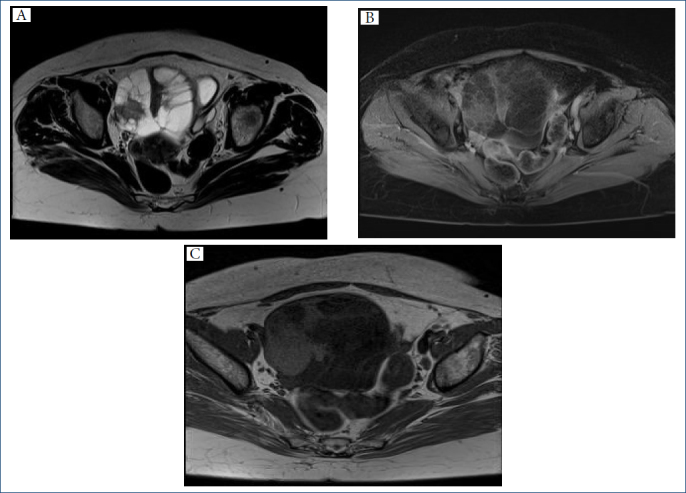
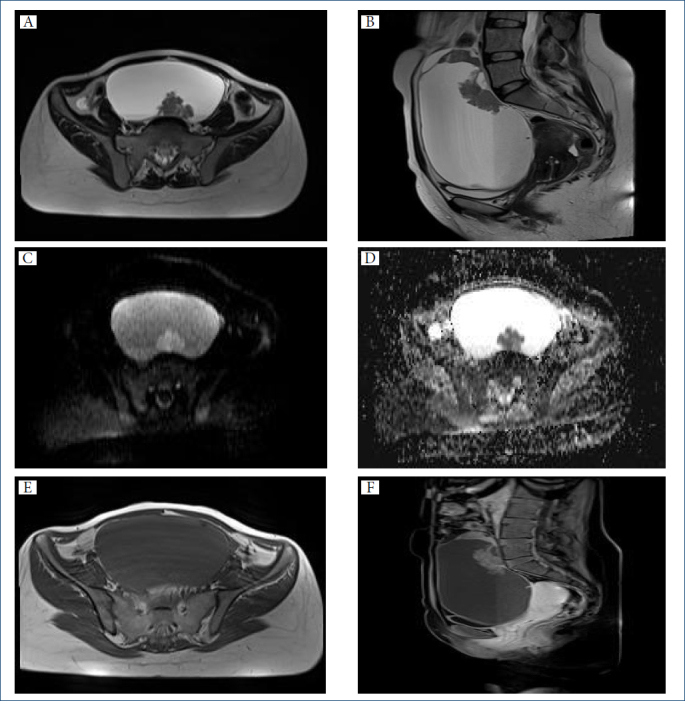
Results: There was no difference between the two groups in terms of maximum tumor size. Age at diagnosis was 56.29±11.92 in the mucinous ovarian carcinoma group and 44.74±13.60 in the mucinous borderline ovarian tumor group (p<0.05). A significant difference was found between the two groups, and it was observed that mucinous borderline ovarian tumors appeared in the younger age group compared to mucinous ovarian carcinomas. Presence of ascites, peritoneal dissemination, lymphadenopathy, and mural nodules was found significantly more frequently in mucinous ovarian carcinomas than in mucinous borderline ovarian tumors. Honeycomb appearance was found more frequently in mucinous borderline ovarian tumor patients than in mucinous ovarian carcinoma patients.
Conclusion: magnetic resonance imaging findings of these two pathologies overlapped considerably. Compared with mucinous borderline ovarian tumors, mucinous ovarian carcinomas frequently had mural nodules larger than 5 mm, larger tumor size, peritoneal dissemination, and abnormal ascites.
Conflict of interest statement
Conflicts of interest: the authors declare there is no conflicts of interest.
Figures
Similar articles
-
Can MRI features differentiate ovarian mucinous carcinoma from mucinous borderline tumor?Eur J Radiol. 2020 Nov;132:109281. doi: 10.1016/j.ejrad.2020.109281. Epub 2020 Sep 12. Eur J Radiol. 2020. PMID: 32961452
-
Salient magnetic resonance imaging findings in the differential diagnosis of benign, borderline and malignant ovarian mucinous tumors.Abdom Radiol (NY). 2025 Feb;50(2):1009-1017. doi: 10.1007/s00261-024-04545-9. Epub 2024 Aug 27. Abdom Radiol (NY). 2025. PMID: 39187694
-
Imaging in gynecological disease (11): clinical and ultrasound features of mucinous ovarian tumors.Ultrasound Obstet Gynecol. 2017 Aug;50(2):261-270. doi: 10.1002/uog.17222. Epub 2017 Jun 22. Ultrasound Obstet Gynecol. 2017. PMID: 28782867
-
Mucinous tumors of the ovary: a review.Int J Gynecol Pathol. 2005 Jan;24(1):4-25. Int J Gynecol Pathol. 2005. PMID: 15626914 Review.
-
Borderline epithelial ovarian tumors: what the radiologist should know.Abdom Radiol (NY). 2021 Jun;46(6):2350-2366. doi: 10.1007/s00261-020-02688-z. Epub 2020 Aug 29. Abdom Radiol (NY). 2021. PMID: 32860524 Review.
Cited by
-
MRI characteristics of ovarian metastasis: differentiation from stomach and colorectal cancer.Jpn J Radiol. 2025 Apr;43(4):676-686. doi: 10.1007/s11604-024-01700-6. Epub 2024 Nov 14. Jpn J Radiol. 2025. PMID: 39538067 Free PMC article.
-
MRI findings of malignant transformation arising from mature cystic teratoma of the ovary: comparison with benign mature cystic teratoma.Jpn J Radiol. 2024 May;42(5):500-507. doi: 10.1007/s11604-023-01521-z. Epub 2023 Dec 26. Jpn J Radiol. 2024. PMID: 38146022 Free PMC article.
-
Prediction of grading of ovarian endometrioid carcinoma using conventional MRI features.Jpn J Radiol. 2025 May;43(5):820-828. doi: 10.1007/s11604-024-01727-9. Epub 2024 Dec 28. Jpn J Radiol. 2025. PMID: 39730935 Free PMC article.
References
-
- Taylor HC. Malignant and semimalignant tumors of the ovary. Surg Gynecol Obstet. 1929;48:204–30.
-
- Riopel MA, Ronnett BM, Kurman RJ. Evaluation of diagnostic criteria and behavior of ovarian intestinal-type mucinous tumors: atypical proliferative (borderline) tumors and intraepithelial, microinvasive, invasive, and metastatic carcinomas. Am J Surg Pathol. 1999;23(6):617–35. doi: 10.1097/00000478-199906000-00001. - DOI - PubMed
MeSH terms
LinkOut - more resources
Full Text Sources
Medical
