Review
. 2023 Oct;53(11):2291-2304.
doi: 10.1007/s00247-023-05709-3. Epub 2023 Jul 19.

Postnatal imaging of conjoined twins: a customized multimodality approach

Affiliations
Review

Postnatal imaging of conjoined twins: a customized multimodality approach

Shaimaa Abdelsattar Mohammad et al. Pediatr Radiol. 2023 Oct.

Abstract

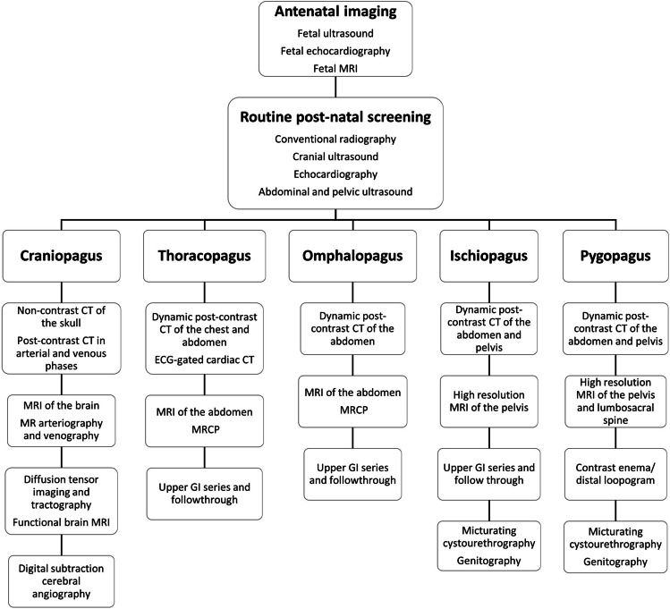
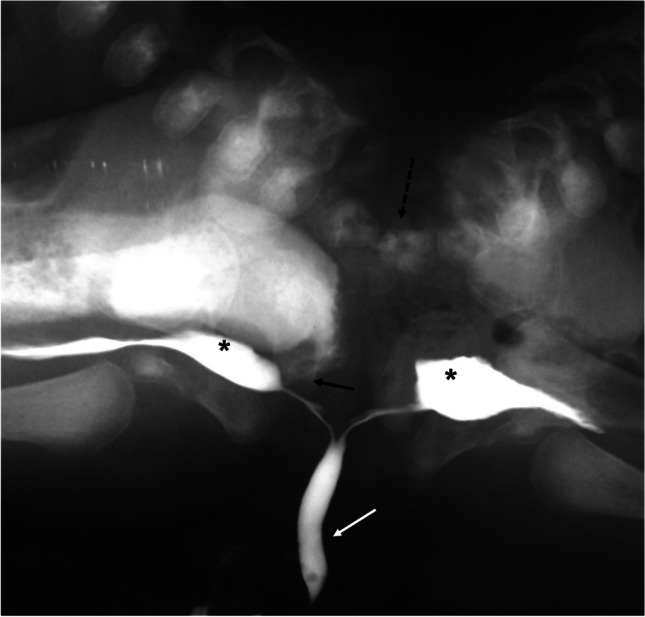
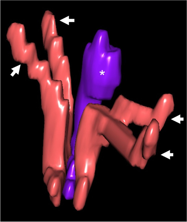
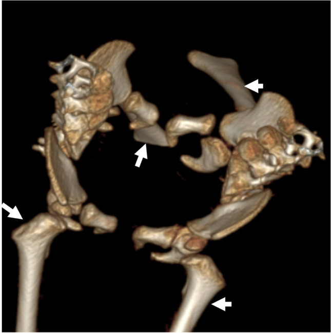
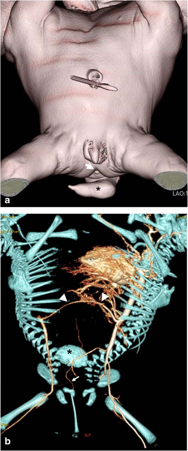
Medical imaging plays a crucial role in the management of conjoined twins. The role of imaging is to explore the anatomy, outline the shared organs and determine whether surgery is feasible. It also serves as a roadmap for successful separation. Additionally, imaging helps with counseling parents about prognosis. This review aims to illustrate recent advances in different imaging modalities and their role in the management of the various types of conjoined twins with an emphasis on relevant tips for optimal imaging.

Keywords: Conjoined twins; Diagnostic imaging; Magnetic resonance imaging; Three-dimensional imaging; Three-dimensional printing; Virtual reality.


Conflict of interest statement

None

Figures

Fig. 1
Fig. 1
15-month-old heteropagus conjoined twin girls. a A photographic images shows the parasitic twin attached to the anterior chest wall of the autositic twin. The parasitic twin has a deformed cranium and incomplete trunk with four limbs. b Maximum intensity projection computed tomography angiography shows an anomalous vessel (arrow) supplying the parasitic twin which originates from the subclavian artery of the autosite. Note the kidney of the parasitic twin (asterisk)
Fig. 2
Fig. 2
A 1-month-old boy presented with abdominal swelling which proved to be a parasitic twin (fetus in fetu). a Coronal reformatted computed tomoagraphy image of the abdomen and pelvis (soft tissue window) of the autosite shows the bony skeleton of the parasitic twin within a fatty mass (arrow). b 3-dimensional reformat of the skeleton nicely demonstrates the presence of a dysplastic sacrum and bones of the pelvis and lower limb at variable degrees of development. c Post-operative photographic image after excision of the parasitic twin
Fig. 3
Fig. 3
16-year-old pygopagus conjoined twin girls. a 3-dimensional (D) surface-rendered image reveals the site of fusion (arrows). Note the colostomy sites in both twins (arrowheads). b 3-D multimask image extracted from a computed tomography (CT) study in the excretory phase shows limited sacral fusion (broken arrow). The right twin has crossed fused kidneys (black arrows), while the left twin has a single kidney with crossed ectopia (white arrow). c CT angiography in the arterial phase (both twins injected simultaneously) shows arterial communication (arrows). d Maximum intensity projection of the coronal fat suppressed T2-weighted magnetic resonance image reveals that both thecal sacs are continuous through a narrow channel (short arrow). Note the single kidney of the left twin (arrow) and crossed fused ectopia of the right twin (arrowheads)
Fig. 4
Fig. 4
Flowchart depicting the sequence of imaging modalities in relatively common types of conjoined twins. CT computed tomography, ECG electrocardiography, GI gastrointestinal, MRCP magnetic resonance cholangiopancreatography, MRI magnetic resonance imaging
Fig. 5
Fig. 5
Magnetic resonance images in 2-year-old craniopagus conjoined twin girls. a Coronal T2-weighted image shows cranial fusion with two separate brains with an intervening common venous sinus (arrow). b Coronal magnetic resonance venography image confirms sharing of the superior sagittal sinus (arrow). c Coronal magnetic resonance arteriography image reveals no significant arterial sharing
Fig. 6
Fig. 6
8-month-old thoracopagus conjoined twin boys. a Clinical photographic image shows thoracic and abdominal fusion with a small omphalocele (arrow). b 3-dimensional reconstruction of the bony skeleton shows sternal fusion (arrow). c Axial computed tomography of the thorax (mediastinal window) following contrast injection into the twin on the right shows opacification of the cardiac chambers of the injected baby only (asterisk), denoting separate cardiac chambers. Note the collapsed right lung with increased density (arrow)
Fig. 7
Fig. 7
Computed tomography (CT) in 4-month-old ischiopagus conjoined twin boys following intravenous contrast injection of each twin on separate days. (ac) are extracted following contrast injection of the right twin, while (d–f) are extracted following contrast injection of the left twin. a, b Maximum intensity projection (MIP) (a) and color-coded 3-dimensional (D) reconstruction) (b) show an abnormal venous channel (arrow) connecting the portal system of both twins with retrograde venous filling into the left twin (arrowhead). c Axial CT image (soft tissue window) with color overlay reveals the lobulated margin of the liver at the site of fusion (arrows). d, e Coronal CT (soft tissue window) (d) and 3-D with color overlay (e) show abnormal venous drainage into the right twin (arrow) after the left twin was injected with contrast. f Axial CT image (soft tissue window) with color overlay shows the liver (black asterisk), spleen (white asterisk), hepatic vein (black arrow) and portal vein (white arrow) of the left twin. (Visible Patient®, Strasbourg, France. Red heart and aorta, cyan hepatic veins, light orange large intestine, rosy brown liver, pink lungs and trachea, dark gray lungs [lower lobes], royal blue portal veins, light pink small intestine, mauve spleen, yellow urinary bladder)
Fig. 8
Fig. 8
Imaging of 4-month-old ischiopagus conjoined twin boys (same patients as in Fig. 7). a Color-coded 3-dimensional (D) image extracted from a delayed phase computed tomography (CT) study. Each baby has two kidneys. There are two urinary bladders (asterisks) seen in the common pelvis. b Axial high resolution-T2-weighted magnetic resonance image with color overlay shows the anatomy of the conjoined pelvis. There are two urinary bladders (asterisks) and a single rectum (#). c Color-coded 3-D CT image of the left twin reveals each ureter draining into a separate bladder (arrows). The left kidney has a duplex collecting system (arrowheads) and proximal ureteric duplication. Note the presence of a hemivertebra (asterisk) (Visible Patient®, Strasbourg, France. Ivory bony skeleton, brown kidneys, pink lungs, tan rectum, orange ureters, violet ureter, yellow urinary bladders)
Fig. 9
Fig. 9
An anteroposterior pelvic radiograph obtained during micturating cystourethrography in 4-week-old pygopagus conjoined twin boys shows a single urethral channel (white arrow) with two separate urinary bladders (asterisks). Note the vertebral fusion (broken arrow) and rectourethral fistula in the right twin (black arrow)
Fig. 10
Fig. 10
4-month-old ischiopagus conjoined twin boys with a common penis (same patients as in Figs. 7 and 8). Magnified 3-diemsnional view of the corporeal tissue extracted from axial high resolution T2-weighted magnetic resonance imaging. There are four corpora cavernosa (two belonging to each twin) (arrows) and a common corpus spongiosum (asterisk). (Visible Patient®, Strasbourg, France)
Fig. 11
Fig. 11
Pelvic fusion of 4-month-old ischiopagus tetrapus conjoined twin boys (same patients as in Figs. 7, 8 and 10). 3-dimensional computed tomography reconstruction of the bony pelvis shows that each baby has two normal lower limbs (arrows)
Fig. 12
Fig. 12
Computed tomography in 17-day-old ischiopagus tripus conjoined twin boys. a Surface-rendered image shows each baby to have one normal leg. There is a third deformed leg (asterisk) attached posteriorly to the conjoined pelvis. Note the single anal orifice (arrow) and single penis and scrotum. b 3-dimensional reconstruction of the skeleton and arterial tree following contrast injection of the left twin reveals a common iliac bone posteriorly (asterisk) and short bones of the deformed limb supplied by a small branch (arrow) arising from a hypoplastic right common iliac artery. A shared superior mesenteric artery (arrowheads) connects the aortas of both twins

Similar articles

Cited by

  • Antenatal imaging diagnosis of thoraco-omphalopagus conjoined twins.
    Guelzim Y, Halfi Mohamed I, Marrakchi S, Krioule S, Tahri S, El Yousfi M, El Hassouni F, Maakoul R, Allali N, Chat L, El Haddad S. Guelzim Y, et al. Radiol Case Rep. 2024 Jun 1;19(8):3478-3482. doi: 10.1016/j.radcr.2024.05.003. eCollection 2024 Aug. Radiol Case Rep. 2024. PMID: 38872747 Free PMC article.
  • Fetal MRI findings in conjoined twin pregnancies.
    Walcutt JE, Kline-Fath BM, Ayyala RS, Lim FY, Nagaraj UD. Walcutt JE, et al. Pediatr Radiol. 2025 Aug 15. doi: 10.1007/s00247-025-06323-1. Online ahead of print. Pediatr Radiol. 2025. PMID: 40815378 Review.

References

    1. Barth RA, Filly RA, Goldberg JD, et al. Conjoined twins: prenatal diagnosis and assessment of associated malformations. Radiology. 1990;177:201–207. doi: 10.1148/radiology.177.1.2204966. - DOI - PubMed
    1. Spencer R. Theoretical and analytical embryology of conjoined twins: part I: embryogenesis. Clin Anat (New York, N.Y.) 2000;13:36–53. doi: 10.1002/(SICI)1098-2353(2000)13:1<36::AID-CA5>3.0.CO;2-3. - DOI - PubMed
    1. Kaufman MH. The embryology of conjoined twins. Childs Nerv Syst. 2004;20:508–525. doi: 10.1007/s00381-004-0985-4. - DOI - PubMed
    1. McHugh K, Kiely EM, Spitz L. Imaging of conjoined twins. Pediatr radiol. 2006;36:899–910. doi: 10.1007/s00247-006-0121-6. - DOI - PubMed
    1. Kingston CA, McHugh K, Kumaradevan J, et al. Imaging in the preoperative assessment of conjoined twins. Radiographics. 2001;21:1187–1208. doi: 10.1148/radiographics.21.5.g01se011187. - DOI - PubMed

LinkOut - more resources