Review
. 2023 Jul 19;9(1):60.
doi: 10.1186/s40798-023-00604-5.

In Vivo Strain Patterns in the Achilles Tendon During Dynamic Activities: A Comprehensive Survey of the Literature

Affiliations
Review

In Vivo Strain Patterns in the Achilles Tendon During Dynamic Activities: A Comprehensive Survey of the Literature

Naomi C Adam et al. Sports Med Open. .

Abstract

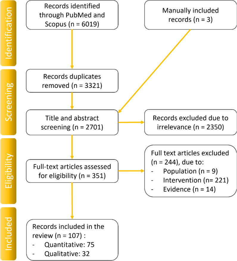
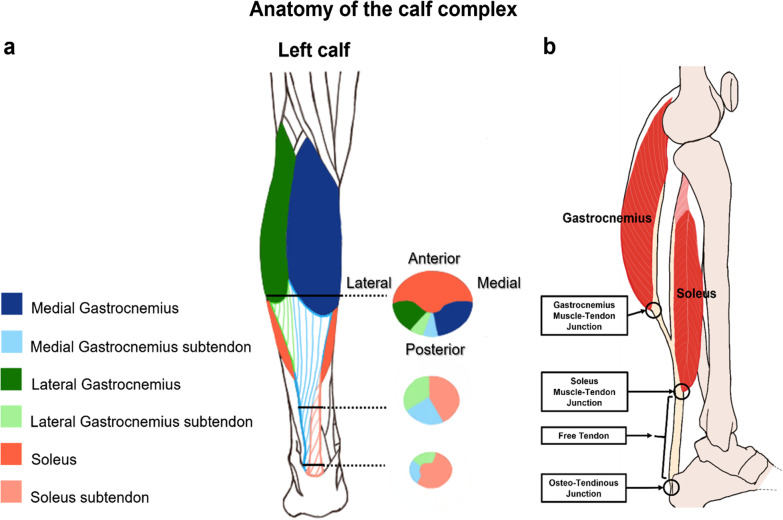
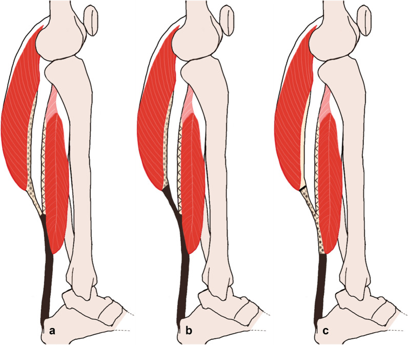
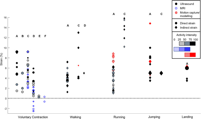
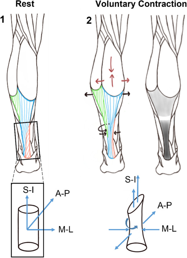
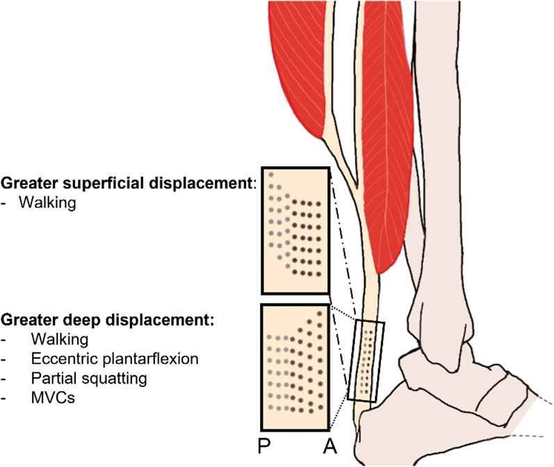
Achilles' tendon (AT) injuries such as ruptures and tendinopathies have experienced a dramatic rise in the mid- to older-aged population. Given that the AT plays a key role at all stages of locomotion, unsuccessful rehabilitation after injury often leads to long-term, deleterious health consequences. Understanding healthy in vivo strains as well as the complex muscle-tendon unit interactions will improve access to the underlying aetiology of injuries and how their functionality can be effectively restored post-injury. The goals of this survey of the literature with a systematic search were to provide a benchmark of healthy AT strains measured in vivo during functional activities and identify the sources of variability observed in the results. Two databases were searched, and all articles that provided measured in vivo peak strains or the change in strain with respect to time were included. In total, 107 articles that reported subjects over the age of 18 years with no prior AT injury and measured while performing functional activities such as voluntary contractions, walking, running, jumping, or jump landing were included in this review. In general, unclear anatomical definitions of the sub-tendon and aponeurosis structures have led to considerable confusion in the literature. MRI, ultrasound, and motion capture were the predominant approaches, sometimes coupled with modelling. The measured peak strains increased from 4% to over 10% from contractions, to walking, running, and jumping, in that order. Importantly, measured AT strains were heavily dependent on measurement location, measurement method, measurement protocol, individual AT geometry, and mechanical properties, as well as instantaneous kinematics and kinetics of the studied activity. Through a comprehensive review of approaches and results, this survey of the literature therefore converges to a united terminology of the structures and their common underlying characteristics and presents the state-of-knowledge on their functional strain patterns.

Keywords: Achilles tendon; Aponeurosis; Dynamic activities; In vivo measurement; Sub-tendon; Tendon strain.


Conflict of interest statement

The authors declare that they have no competing interests.

Figures

Fig. 1
Fig. 1
PRISMA diagram of the articles included in this review
Fig. 2
Fig. 2
a Three axial cross sections of the AT sub-tendons demonstrating their natural twist [19], b sagittal cross section indicating common anatomic naming conventions of the triceps surae muscles and the Achilles tendon (note: spaces between soft tissues are exaggerated for graphical clarity)
Fig. 3
Fig. 3
Three different definitions ac of the gastrocnemii aponeuroses (dotted), soleus aponeurosis (lines), and Achilles/free tendon (solid black) are found in the literature; however, in this review convention “c” was chosen because it is most commonly used (note: spaces between soft tissues are exaggerated for graphical clarity)
Fig. 4
Fig. 4
Peak strain results across activities: results are split by Achilles tendon location. The size of the data points is proportional to the number of participants. Colours indicate measurement methods; the filling gradient represents the speed or intensity of the activity and the shapes if the strain had been directly measured or was an indirectly obtained value. A: Medial gastrocnemius sub-tendon, B: lateral gastrocnemius sub-tendon, C: free tendon, D: medial gastrocnemius aponeurosis, E: lateral gastrocnemius aponeurosis, F: soleus aponeurosis
Fig. 5
Fig. 5
Strain patterns of the Achilles tendon during a voluntary contraction. The two cylinders represent a close-up of the free tendon at rest (panel 1) and during contraction (panel 2), assuming no change in knee and ankle joint angles. In panel 2, the red arrows indicate the shortening and bulging of the gastrocnemius muscle during contraction, while the black arrows illustrate the positive transversal strain (widening) around the gastrocnemius aponeurosis, as well as the negative transverse strain (thinning) at the free tendon. Additionally, the circular black arrow around the free tendon represents the rotation undergone during contraction. The grey scale scheme represents the magnitude of the longitudinal strain undergone by the tissue: the darker the colour, the greater the longitudinal strain. The white region around the soleus muscle tendon junction indicates no strain or even a negative longitudinal strain [86]. (S-I: superior–inferior, A-P: anterior–posterior, M-L: medio–lateral)
Fig. 6
Fig. 6
Sagittal cut of the free tendon showing the difference in longitudinal elongation between superficiaol and deep layers during several activities. The grey dots represent the superficial/gastrocnemius layers, and the black dots represent the deep/soleus layer. A and P stand for the anterior and posterior directions along the sagittal plane

References

    1. Scott SH, Winter DA. Internal forces of chronic running injury sites. Med Sci Sports Exerc. 1990;22(3):357–369. doi: 10.1249/00005768-199006000-00013. - DOI - PubMed
    1. Gross CE, Nunley JA., 2nd Acute Achilles tendon ruptures. Foot Ankle Int. 2016;37(2):233–239. doi: 10.1177/1071100715619606. - DOI - PubMed
    1. Jarvinen TA, Kannus P, Maffulli N, Khan KM. Achilles tendon disorders: etiology and epidemiology. Foot Ankle Clin. 2005;10(2):255–266. doi: 10.1016/j.fcl.2005.01.013. - DOI - PubMed
    1. Jozsa L, Kannus P. Histopathological findings in spontaneous tendon ruptures. Scand J Med Sci Sports. 1997;7(2):113–118. doi: 10.1111/j.1600-0838.1997.tb00127.x. - DOI - PubMed
    1. Maffulli N, Sharma P, Luscombe KL. Achilles tendinopathy: aetiology and management. J R Soc Med. 2004;97(10):472–476. doi: 10.1177/0141076809701004. - DOI - PMC - PubMed

LinkOut - more resources