. 2023 Jul 19;18(7):e0288810.
doi: 10.1371/journal.pone.0288810. eCollection 2023.

Molecular insights into the inhibitory potential of anthocyanidins on glucokinase regulatory protein

Affiliations

Molecular insights into the inhibitory potential of anthocyanidins on glucokinase regulatory protein

Christian Kenneth et al. PLoS One. .

Abstract

Computational methods were used to investigate six anthocyanidins exhibiting antidiabetic activity by inhibiting glucokinase regulatory protein (GKRP) activity. Density functional theory was used to optimise the geometry of anthocyanidins and calculate their quantum chemical properties. A blind docking method was employed to conduct a molecular docking study, which revealed that delphinidin (Del), cyanidin (Cya), and pelargonidin (Pel) as potential GKRP inhibitors with the lowest binding free energy of -8.7, -8.6, and -8.6 kcal/mol, corresponding to high binding affinity. The molecular dynamics study further verified the blind docking results by showing high GKRP-F1P complex stability and high binding affinity calculated through the MM/GBSA method, upon the binding of pelargonidin. The lower RMSF values of pivotal GK-interacting residues for GKRP-F1P-Pel compared to GKRP-F1P, as a positive control, indicating pelargonidin ability to maintain the inactive conformation of GKRP through the inhibition of GK binding. The key residues that control the binding of the F1P to GKRP and anthocyanidin to GKRP-F1P were also identified in this study. Altogether, pelargonidin is anthocyanidins-derived natural products that have the most potential to act as inhibitors of GKRP and as antidiabetic nutraceuticals.


Conflict of interest statement

The authors have declared that no competing interests exist.

Figures

Fig 1
Fig 1. Illustration in silico method to analyse the antidiabetic activity of anthocyanidins against GKRP.
Fig 2
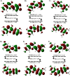
Fig 2
HOMO-LUMO diagram of anthocyanidins at pH 7: (A) Cya, (B) Del, (C) Mal, (D) Pel, (E) Peo, and (F) Pet with HOMO-LUMO gap (HLG) depicted with GaussView 6.
Fig 3
Fig 3. Molecular electrostatic potential (MEP) map of each anthocyanidin illustrated using VMD.
Fig 4
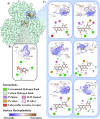
Fig 4
(A) The binding site of anthocyanidins and F1P within the GKRP, types and distance of non-covalent interactions with interacting residual surface hydrophobicity of (B) F1P-GKRP and (C) anthocyanidins-GKRP: first row from left: Cya and Del, second row from left: Mal and Pel, third row from left: Peo and Pet; are visualised with Protein Imager and Discovery Studio Visualizer.
Fig 5
Fig 5
(A) RMSD and (B) cluster distribution of the GKRP backbone for the triplicates of the simulated GKRP-F1P and GKRP-F1P-anthocyanidin systems.
Fig 6
Fig 6
(A-C) RMSF graph of GKRP backbone residues in triplicates, with residues highlighted in gray and (B) brown consecutively indicating the crucial residues of the establishment GKRP-F1P and (C) GKRP-GK complex.
Fig 7
Fig 7
(A) Time-dependent Rg trajectories and (B) SASA dynamics of GKRP for the simulated GKRP-F1P and GKRP-F1P-anthocyanidin systems in triplicates.
Fig 8
Fig 8. Gibbs free energy landscape of GKRP structure plotted between RMSD and Rg of 100 ns MD simulation for each system and replicas.
Fig 9
Fig 9
Box plot visualisation: (A) the MM/GBSA calculation of the binding energy of GKRP-F1P and (B) GKRP-anthocyanidin from the three replicates.
Fig 10
Fig 10. Decomposition plot (MM/GBSA) of amino acid residues within 5 Å of F1P binding site with their contributing interaction energy from each system replica.
Shades of orange: F1P, shades of red: Cya, shades of green: Del, shades of blue: Pel; from above to below: #1 to #3.
Fig 11
Fig 11. Decomposition diagram (MM/GBSA) of amino acid residues within 5 Å of anthocyanidin binding site with their contributing interaction energy from each system replica.
Shades of red: Cya, shades of green: Del, shades of blue: Pel; from above to below: #1 to #3.

References

    1. Goyal R, Jialal I. Diabetes Mellitus Type 2. StatPearls. Treasure Island: StatPearls Publishing; 2022.
    1. Chaudhury A, Duvoor C, Reddy Dendi VS, Kraleti S, Chada A, Ravilla R, et al.. Clinical Review of Antidiabetic Drugs: Implications for Type 2 Diabetes Mellitus Management. Front Endocrinol (Lausanne). 2017;8. doi: 10.3389/fendo.2017.00006 - DOI - PMC - PubMed
    1. Fang J. Bioavailability of anthocyanins. Drug Metab Rev. 2014;46: 508–520. doi: 10.3109/03602532.2014.978080 - DOI - PubMed
    1. Tian L, Tan Y, Chen G, Wang G, Sun J, Ou S, et al.. Metabolism of anthocyanins and consequent effects on the gut microbiota. Crit Rev Food Sci Nutr. 2019;59: 982–991. doi: 10.1080/10408398.2018.1533517 - DOI - PubMed
    1. Oliveira H, Fernandes A, Brás NF, Mateus N, de Freitas V, Fernandes I. Anthocyanins as antidiabetic agents—in vitro and in silico approaches of preventive and therapeutic effects. Molecules. 2020;25: 1–30. doi: 10.3390/molecules25173813 - DOI - PMC - PubMed

Publication types

MeSH terms