. 2023 Jul 19;21(7):e3002112.
doi: 10.1371/journal.pbio.3002112. eCollection 2023 Jul.

Targeting AAV vectors to the central nervous system by engineering capsid-receptor interactions that enable crossing of the blood-brain barrier

Affiliations

Targeting AAV vectors to the central nervous system by engineering capsid-receptor interactions that enable crossing of the blood-brain barrier

Qin Huang et al. PLoS Biol. .

Abstract

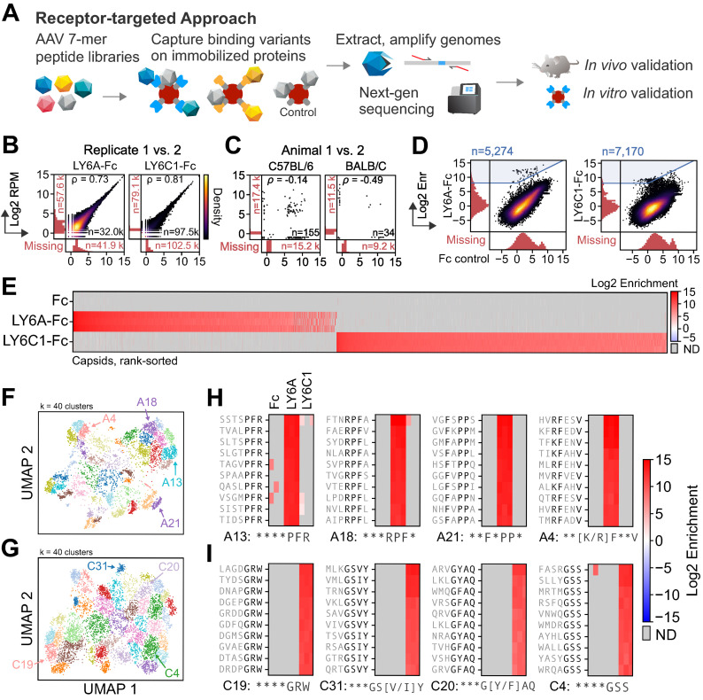
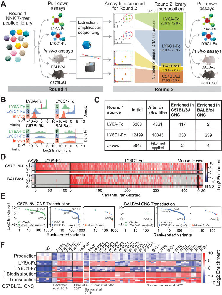
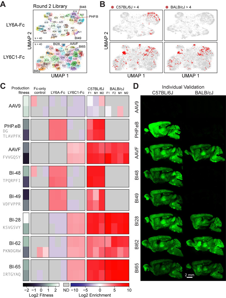
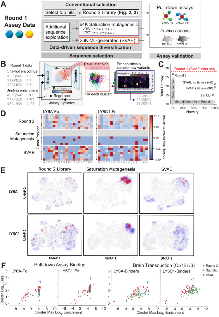
Viruses have evolved the ability to bind and enter cells through interactions with a wide variety of cell macromolecules. We engineered peptide-modified adeno-associated virus (AAV) capsids that transduce the brain through the introduction of de novo interactions with 2 proteins expressed on the mouse blood-brain barrier (BBB), LY6A or LY6C1. The in vivo tropisms of these capsids are predictable as they are dependent on the cell- and strain-specific expression of their target protein. This approach generated hundreds of capsids with dramatically enhanced central nervous system (CNS) tropisms within a single round of screening in vitro and secondary validation in vivo thereby reducing the use of animals in comparison to conventional multi-round in vivo selections. The reproducible and quantitative data derived via this method enabled both saturation mutagenesis and machine learning (ML)-guided exploration of the capsid sequence space. Notably, during our validation process, we determined that nearly all published AAV capsids that were selected for their ability to cross the BBB in mice leverage either the LY6A or LY6C1 protein, which are not present in primates. This work demonstrates that AAV capsids can be directly targeted to specific proteins to generate potent gene delivery vectors with known mechanisms of action and predictable tropisms.


Conflict of interest statement

I have read the journal’s policy and the authors of this manuscript have the following competing interests: BED is a scientific founder at Apertura Gene Therapy and a scientific advisory board member at Tevard Biosciences. BED, QH, KYC, and FEE are named inventors on patent applications filed by the Broad Institute of MIT and Harvard related to this study. Remaining authors declare that they have no competing interests.

Figures

Fig 1
Fig 1. In vitro pull-down assays yield capsids that selectively bind LY6A or LY6C1.
(A) A capsid library is screened for the ability to bind immobilized target Fc-fusion proteins. Bound capsid sequences are extracted and subjected to NGS. Hits are incorporated into a focused library for in vivo and in vitro validation. (B, C) The Pearson correlation of the log2 normalized read count (RPM) are shown between biological replicates (n = 3, only 1 pair shown) (B) and between animals (n = 2) (C). Variants detected in 1 replicate or animal and not the other are shown in the marginal histograms. (D) The variant log2 enrichment (average RPM between replicates, normalized to the starting library RPM) plotted between the target (y-axis) and Fc-only control (x-axis) shows a majority of variants with nonspecific binding and a minority (blue highlighted region) with target-specific binding. The variants detected in 1 assay and not the other are shown in the marginal histograms. (E) The log2 enrichment of the selected variants highlighted in blue in (D) with each replicate’s enrichment plotted in separate rows (n = 3). ND = not detected. (F, G) The sequences that bound LY6A (F) or LY6C1 (G) from Libraries 1 and 2 were one-hot encoded, jointly projected with UMAP, and jointly clustered with a Gaussian mixture model (k = 40, S1–S6 Data). (H, I) Four clusters for each target from (F, G) were manually selected based on whether there was a clear motif 2–4 amino acids in length that matched either existing reference sequence (LY6A-binding: ***PFR, ***RPF, LY6C1-binding: ***G[Y/F]AQ) or represented a motif not yet seen in published studies. Consensus motifs are defined per-position, with flexible amino acid residues (asterisks) and fixed residues (present in more than 40% of the cluster’s sequences; black letters). The underlying data supporting Fig 1B and 1D and 1E can be found at https://doi.org/10.5281/zenodo.7689794: library1.csv; Fig 1C at https://doi.org/10.5281/zenodo.7689794: library2_invivo.csv; Fig 1F and 1H at https://doi.org/10.5281/zenodo.7689794: LY6A_joint_umap_l1_l2.csv; Fig 1G and 1I at https://doi.org/10.5281/zenodo.7689794: LY6C1_joint_umap_l1_l2.csv. NGS, next-generation sequencing; RPM, reads per million.
Fig 2
Fig 2. Round 2 validation of LY6A- and LY6C1-binding variants identified thousands of capsids with CNS transduction activity.
(A) The Round 2 library was composed of a selection of top performing variants from the Round 1 assays for LY6A binding, LY6C1 binding, in vivo CNS transduction, and published reference sequences. The Round 2 library was subjected to screening as in Round 1. (B) The distributions show the Round 2 library performance in the pull-down assays and the CNS transduction of BALB/cJ and C57BL/6J mice, grouped and colored by the Round 1 selection source of each variant. The red lines indicate the thresholds set for the filters applied in (C). (C) The hits in Round 2 were identified as follows: target-binding capsids from the Round 1 screen were first filtered on their respective target binding activity in the Round 2 screen (LY6A: log2 enrichment > 0, LY6C1: log2 enrichment > -2). Variants were then filtered on Round 2 in vivo CNS transduction (log2 enrichment > 4 in either mouse strain and detected in at least 2 animals within that strain). (D) The in vivo log2 enrichment scores in C57BL/6J and BALB/cJ mice of Round 2 library variants that were filtered for high in vivo log2 enrichment scores in (C). The scores in individual animals (M*, F*) for each strain are shown alongside the average across animals (all). Variants are shown grouped and colored by their Round 1 selection source and rank-sorted on a combined score of C57BL/6J and BALB/cJ transduction. (E) The filtered variants from (C) are shown grouped and colored by their Round 1 selection source and rank-sorted separately for each mouse strain. Reference controls and AAV9 are marked with crosses. Variants identified in both the Round 1 pull-down assays and in vivo screen are displayed as filled dots. (F) The target binding and C57BL/6J CNS biodistribution or transduction phenotypes of reference capsids with CNS tropisms are shown. Each capsid is represented by at least two 7-mer AA replicates (each column indicates a separate replicate). The underlying data supporting Fig 2A can be found at https://doi.org/10.5281/zenodo.7689794: round2_codons_separate.csv; Fig 2B–E at https://doi.org/10.5281/zenodo.7689794: round2_codons_merged.csv; Fig 2F at https://doi.org/10.5281/zenodo.7689794: SVAE_SM_library_references_only.csv. AA, amino acid; AAV, adeno-associated virus; CNS, central nervous system.
Fig 3
Fig 3. LY6A- and LY6C1-binding capsids identified in the pull-down assays cross the mouse BBB.
(A) The UMAPs of Round 2 library variants are shown projected onto the UMAPs of Round 1 variants. Variant sequences were clustered with K-means (LY6A, k = 25; LY6C1, k = 30) (see cluster summaries in S10 Data). (B) The Round 2 variants with an in vivo brain transduction log2 enrichment of > 4 in C57BL/6J mice (left) and BALB/cJ mice (right) are marked in red. (C) The Round 2 in vivo screen results for the reference capsids and 5 Round 2 variants selected for individual characterization are shown. Each variant is represented by two 7-mer AA replicates indicated by separate rows. ND = not detected. (D) Representative brain images are shown for the capsids in (C) that were individually tested in C57BL/6J mice (left) and BALB/cJ mice (right). The underlying data supporting Fig 3A and 3B can be found at https://doi.org/10.5281/zenodo.7689794: round2_codons_merged.csv; Fig 3C at https://doi.org/10.5281/zenodo.7689794: round2_codons_separate.csv. AA, amino acid; BBB, blood–brain barrier.
Fig 4
Fig 4. A single round of screening data can be used with SVAE and saturation mutagenesis to generate additional functional sequences.
(A) Round 1 data were used to explore additional sequence diversity via 2 methods: saturation mutagenesis around 2 motifs (LY6A ***[K/R]PF[I/L], LY6C1 ***G[W/Y]S[A/S]) and SVAE ML generation. (B) The SVAE was trained on Round 1, library 1 sequences (encoder/decoder blocks) and binding enrichments (regression block). During training, these blocks were jointly optimized. High-binding enrichment sequences were isolated and re-clustered, and new sequences were sampled from each cluster’s position weight matrix (PWM) (S12 Fig and Materials and methods). (C) The total statistical entropy (summed entropies across all 7 amino acid positions) versus novelty (the fraction not found in Round 1) of each set of variants is shown. (D) Amino acid frequencies relative to uniform (1/20 chance of each) for the indicated libraries’ LY6A-Fc (left) and LY6C1-Fc binders (right). (E) The UMAP projection of sequence exploration for LY6A- (top row) and LY6C1-binders (bottom row) are mapped onto the same UMAP projection as Figs 1–3; the Round 1 UMAP is reproduced in gray in each plot. Sequences with a log2 enrichment for production fitness > -1.0 (blue) and both fitness > -1.0 and in vivo log2 enrichment of > 3 (red) are shown for the Round 2 library (left), saturation mutagenesis (center), and SVAE (right). (F) Each point represents a cluster from (E), using the same cluster boundaries as in Fig 1F and 1G, plotted by cluster size versus the cluster’s maximum log2 enrichment in the binding or transduction assay. Log2 enrichments were calibrated using control sequences (S11 Fig, S12–S23 Data); no calibration adjustment exceeded 2.0. The underlying data supporting Fig 4 can be found at https://doi.org/10.5281/zenodo.7689794: round2_codons_merged.csv and at https://doi.org/10.5281/zenodo.7689794: SVAE_SM_library_codons_merged.csv. ML, machine learning; SVAE, supervised variational auto-encoder.

References

    1. Chan KY, Jang MJ, Yoo BB, Greenbaum A, Ravi N, Wu W-L, et al. Engineered AAVs for efficient noninvasive gene delivery to the central and peripheral nervous systems. Nat Neurosci. 2017;20:1172–1179. doi: 10.1038/nn.4593 - DOI - PMC - PubMed
    1. Deverman BE, Pravdo PL, Simpson BP, Kumar SR, Chan KY, Banerjee A, et al. Cre-dependent selection yields AAV variants for widespread gene transfer to the adult brain. Nat Biotechnol. 2016;34:204–209. doi: 10.1038/nbt.3440 - DOI - PMC - PubMed
    1. Nonnenmacher M, Wang W, Child MA, Ren X-Q, Huang C, Ren AZ, et al. Rapid evolution of blood-brain-barrier-penetrating AAV capsids by RNA-driven biopanning. Mol Ther Methods Clin Dev. 2021;20:366–378. doi: 10.1016/j.omtm.2020.12.006 - DOI - PMC - PubMed
    1. Krolak T, Chan KY, Kaplan L, Huang Q, Wu J, Zheng Q, et al. A high-efficiency AAV for endothelial cell transduction throughout the central nervous system. Nature Cardiovascular Research. 2022;1:389–400. doi: 10.1038/s44161-022-00046-4 - DOI - PMC - PubMed
    1. Körbelin J, Dogbevia G, Michelfelder S, Ridder DA, Hunger A, Wenzel J, et al. A brain microvasculature endothelial cell-specific viral vector with the potential to treat neurovascular and neurological diseases. EMBO Mol Med. 2016;8:609–625. doi: 10.15252/emmm.201506078 - DOI - PMC - PubMed

Substances