. 2023 Jul 21;9(29):eadf7826.
doi: 10.1126/sciadv.adf7826. Epub 2023 Jul 19.

Mechanisms of nuclear pore complex disassembly by the mitotic Polo-like kinase 1 (PLK-1) in C. elegans embryos

Affiliations

Mechanisms of nuclear pore complex disassembly by the mitotic Polo-like kinase 1 (PLK-1) in C. elegans embryos

Sylvia Nkombo Nkoula et al. Sci Adv. .

Abstract

The nuclear envelope, which protects and organizes the genome, is dismantled during mitosis. In the Caenorhabditis elegans zygote, nuclear envelope breakdown (NEBD) of the parental pronuclei is spatially and temporally regulated during mitosis to promote the unification of the maternal and paternal genomes. Nuclear pore complex (NPC) disassembly is a decisive step of NEBD, essential for nuclear permeabilization. By combining live imaging, biochemistry, and phosphoproteomics, we show that NPC disassembly is a stepwise process that involves Polo-like kinase 1 (PLK-1)-dependent and -independent steps. PLK-1 targets multiple NPC subcomplexes, including the cytoplasmic filaments, central channel, and inner ring. PLK-1 is recruited to and phosphorylates intrinsically disordered regions (IDRs) of several multivalent linker nucleoporins. Notably, although the phosphosites are not conserved between human and C. elegans nucleoporins, they are located in IDRs in both species. Our results suggest that targeting IDRs of multivalent linker nucleoporins is an evolutionarily conserved driver of NPC disassembly during mitosis.


Figures
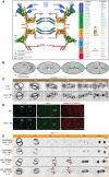
Fig. 1.
Fig. 1.. PLK-1 triggers nuclear pore complexes disassembly independently of lamina depolymerization.
(A) Schematic of the nuclear pore complex and nomenclature of the NUPs in human cells and in C. elegans (NPP). (B) After meeting of the female (pink) and male (blue) pronuclei at the posterior pole, the nucleo-centrosomal complex undergoes a 90° rotation to align along the anteroposterior axis of the embryo. Parental chromosomes congress on the metaphase plate before the initiation of anaphase. In telophase, the NE reforms around decondensing chromosomes. During mitosis, remnant NE (yellow) remains around the segregating chromosomes. NPC, nuclear pore complex; NE/ER, nuclear envelope/endoplasmic reticulum. (C) Micrographs of wild-type (WT) or lmn-1 8A mutant embryos expressing GFP::NPP-22NDC1 in mitosis. The green arrow marks the presence of a membrane gap in control WT as opposed to lmn-1 8A embryos (red arrow). Timings in seconds are relative to pronuclei juxtaposition (0 s). The anterior is to the left in this and other figures. The asterisk (*) indicates the NUPs that are endogenously tagged in this and other figures. All panels are at the same magnification. Scale bars, 3 μm. n is the number of embryos analyzed in this and other figures. Data were collected from three independent experiments. (D) Confocal images of fixed WT and lmn-1 8A mutant two-cell embryos stained with LMN-1 (green) and Mab414 (red) antibodies and counterstained with 4′,6-diamidino-2-phenylindole (DAPI) (blue). All panels are at the same magnification. Scale bar, 10 μm. (E) Micrographs of live WT or lmn-1 8A embryos expressing GFP::NPP-19NUP53 in mitosis exposed to control, or plk-1(RNAi). The red arrowheads point to GFP::NPP-19NUP53 persisting on the NE during mitosis. Timings in seconds are relative to pronuclei juxtaposition (0 s). All panels are at the same magnification. Scale bars, 3 μm. Data were collected from three independent experiments.
Fig. 2.
Fig. 2.. Nucleoporins of the cytoplasmic filaments, the central channel, and the inner ring accumulate at the nuclear envelope upon plk-1 inactivation.
(A to F) Micrographs of embryos expressing the indicated tagged NUPs exposed to control or plk-1(RNAi) starting when the pronuclei are juxtaposed, centered, and aligned along the anteroposterior axis of the embryo (time 0). The red arrowheads show the persisting NUPs on the NE during mitosis. All panels are at the same magnification. Scale bar, 3 μm. Data were collected from three independent experiments.
Fig. 3.
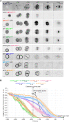
Fig. 3.. Timing of NPC subcomplexes disassembly in the C. elegans zygote relative to the rupture of the nuclear permeability barrier.
(A) Micrographs of one-cell embryos expressing PLK-1::sGFP or the indicated tagged NUPs from pronuclear migration and meeting to anaphase onset (time 0). All panels are at the same magnification. Scale bar, 10 μm. (B) Graph presenting the quantification of GFP::NPP signal intensity at the NE in the C. elegans zygote from pronuclear nuclear migration to anaphase onset (time 0). The average signal intensity of the fluorescently tagged NPPs at the NE 350 s before anaphase was arbitrarily defined as 1. The means ± SEM is presented for n = 6 embryos. Data were collected from three independent experiments. The dark line presents the quantification of the nuclear permeability barrier starting 200 s before anaphase onset. a.u., arbitrary units.
Fig. 4.
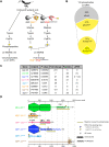
Fig. 4.. A biochemical and phosphoproteomic screen for PLK-1 targets at the nuclear pore complexes.
(A) Flowchart of the approach used to map the NUPs phosphoproteome and identify NUPs specifically binding the Plk1 PBD. Embryonic extracts prepared from young adults were digested with trypsin and phosphopeptides were affinity-purified using FeNTA immobilized metal ion affinity chromatography columns before identification by tandem mass spectrometry (MS/MS). A fraction of the embryonic extracts were incubated on an affinity matrix consisting of the glutathione S-transferase (GST)–PBD WT or mutated on the phosphopincers. After several washing steps, retained proteins were digested with trypsin and phosphopeptides were affinity-purified on FeNTA before identification by liquid chromatography–MS/MS (LC-MS/MS). (B) Circle chart showing the proportion of phosphorylation sites identified on NUPs presenting the consensus for phosphorylation by proline-directed kinases [(pS/pT-P), Cyclin-Cdk]. The circle chart below presents the percentage of these phosphosites matching the consensus for non–self-priming phosphorylation (S-pS/pT-P). (C) Table summarizing the label-free quantitative MS analysis of the GST-PBD WT or mutated on the phosphopincers pull-downs from three independent C. elegans embryonic extracts. The P value (<0.05) indicates statistical significance for the enrichment of the NUPs in the GST-PBD pull-down. pPDS (phosphorylated Polo-docking sites) indicate the number of Polo-docking sites identified in the different NUPs. The value in parentheses corresponds to cases where a phosphorylated peptide contains a Polo-docking site but the exact position of the phosphorylation is ambiguous. (D) Domain organization and distribution of the phosphorylation sites on the NUPs specifically retained on the GST-PLK-1 PBD affinity matrix: MEL-28ELYS, NPP-19NUP53, NPP-10NNUP98, NPP-14NUP214, NPP-1NUP54, and NPP-12gp210. CBD, chromatin-binding domain; RRM, RNA recognition motif; APD, auto-proteolytic domain; CC, coiled-coiled.
Fig. 5.
Fig. 5.. Phosphorylated NPP-10NNUP98 binds the PLK-1 PBD and contributes to PLK-1 recruitment to the nuclear envelope.
(A) Micrographs of an embryo expressing PLK-1::sGFP (shown alone and in green in the merged images) and mCherry::HIS-11 (magenta, in the merged image) exposed to control (ctrl), npp-14NUP214, npp-10NNUP98, npp-19NUP53, or npp-1NUP54 RNAi. All panels are at the same magnification. Scale bar, 10 μm. The graph on the right presents the quantification of PLK-1::sGFP at the NE in the different RNAi conditions 200 s before anaphase onset. **P < 0.01 and ****P < 0.0001; ns, not significant. Data were collected from three independent experiments. (B and C) Micrographs of embryos expressing NPP-1NUP54::GFP (B) or GFP::NPP-11NUP62 (C) exposed to control (ctrl), npp-10NNUP98, or npp-19NUP53 RNAi. The graphs on the right present the quantification of NPP-1NUP54::GFP or GFP::NPP-11NUP62 average intensity at the NE at six time points relative to anaphase onset (timing in seconds is relative to anaphase onset). *P < 0.05 and ****P < 0.0001. Data were collected from three independent experiments. (D) Schematic and domain organization of NPP-10NNUP98. The C-terminal disordered NPP-10NNUP98 (513 to 821) fragment contains three Polo-docking sites matching the consensus for non–self-priming (yellow, phosphorylatable by Cyclin-Cdk1) and five sites matching the consensus for self-priming (blue, phosphorylatable by PLK-1). The sites that are circled have been identified in vivo. Besides the WT, two additional fragments containing the three non–self-priming Polo-docking sites (3A), or all the Polo-docking sites substituted with alanine are presented. (E and F) In vitro kinase assays were performed with Cyclin B–Cdk1 or PLK-1 kinases and the NPP-10NNUP98 (513 to 821) fragments WT, 3A, or 8A tagged with the maltose-binding protein (MBP) as substrates. The samples were subjected to SDS–polyacrylamide gel electrophoresis (PAGE), followed by a far-Western ligand-binding assay using the PBD fused to GST (top). The bottom panel shows the Stain-Free Blot (Chemidoc, Bio-Rad) of the same membrane.
Fig. 6.
Fig. 6.. Multisite phosphorylation of NPP-19NUP53 is required for the disassembly of the inner ring complex.
(A) NPP-19NUP53 contains three Polo-docking sites matching the consensus for non–self-priming (yellow) and seven sites matching the consensus for self-priming (blue). Besides the WT, two additional NPP-19NUP53 variants containing the three non–self-priming Polo-docking sites (3A) or all the Polo-docking sites (10A) substituted with alanine are presented. The sites that are circled have been identified in vivo. (B) In vitro kinase assays were performed with Cyclin B–Cdk1 or PLK-1 kinases and the NPP-19NUP53 WT, 3A, or 10A tagged with the GST as substrates. The samples were subjected to SDS-PAGE, followed by a far-Western ligand-binding assay using the PBD fused to GST (top). The bottom panel shows the Stain-Free Blot (Chemidoc, Bio-Rad) of the same membrane. (C to E) Micrographs of control (top) or npp-19NUP53 10A mutant embryos (bottom) expressing NPP-1NUP54::GFP (C), GFP::NPP-13NUP93 (D), or GFP::NPP-8NUP155 (E) during mitosis. Timings in seconds are relative to pronuclei juxtaposition (0). All panels are at the same magnification. Scale bar, 3 μm. Data were collected from three independent experiments.
Fig. 7.
Fig. 7.. PLK-1 is recruited to multiple multivalent nucleoporins to dismantle the nuclear pore complexes.
Dynamics of NPCs during the first mitotic division of the C. elegans embryo. During pronuclear migration and meeting, the Y-complex and nuclear basket NUPs leave the NPC. PLK-1 is then recruited to the NPC, 250 s before anaphase onset, via its PBD domain in a phospho-dependent manner just before NEBD when the two pronuclei are juxtaposed. PLK-1 is recruited via the multivalent NUPs NPP-10NNUP98, NPP-19NUP53, the central channel NUPs, and by NPP-14NUP214 phosphorylated at Polo-docking sites by Cyclin B–Cdk1 and PLK-1 itself. PLK-1 recruitment and docking on NUPs promote NPC disassembly.

Update of

Similar articles

Cited by

References

    1. L. Champion, M. I. Linder, U. Kutay, Cellular reorganization during mitotic entry. Trends Cell Biol. 27, 26–41 (2017). - PubMed
    1. P. De Magistris, W. Antonin, The dynamic nature of the nuclear envelope. Curr. Biol. 28, R487–R497 (2018). - PubMed
    1. B. Hampoelz, A. Andres-Pons, P. Kastritis, M. Beck, Structure and assembly of the nuclear pore complex. Annu. Rev. Biophys. 48, 515–536 (2019). - PubMed
    1. D. H. Lin, A. Hoelz, The structure of the nuclear pore complex (an update). Annu. Rev. Biochem. 88, 725–783 (2019). - PMC - PubMed
    1. E. Dultz, M. Wojtynek, O. Medalia, E. Onischenko, The nuclear pore complex: Birth, life, and death of a cellular Behemoth. Cell 11, 1456 (2022). - PMC - PubMed

Publication types

MeSH terms

Substances