. 2023 Aug 2;15(30):35973-35985.
doi: 10.1021/acsami.3c06633. Epub 2023 Jul 19.

Semiconducting Polymer Nanoporous Thin Films as a Tool to Regulate Intracellular ROS Balance in Endothelial Cells

Affiliations

Semiconducting Polymer Nanoporous Thin Films as a Tool to Regulate Intracellular ROS Balance in Endothelial Cells

Miryam Criado-Gonzalez et al. ACS Appl Mater Interfaces. .

Abstract

The design of soft and nanometer-scale photoelectrodes able to stimulate and promote the intracellular concentration of reactive oxygen species (ROS) is searched for redox medicine applications. In this work, we show semiconducting polymer porous thin films with an enhanced photoelectrochemical generation of ROS in human umbilical vein endothelial cells (HUVECs). To achieve that aim, we synthesized graft copolymers, made of poly(3-hexylthiophene) (P3HT) and degradable poly(lactic acid) (PLA) segments, P3HT-g-PLA. In a second step, the hydrolysis of sacrificial PLA leads to nanometer-scale porous P3HT thin films. The pore sizes in the nm regime (220-1200 nm) were controlled by the copolymer composition and the structural arrangement of the copolymers during the film formation, as determined by atomic force microscopy (AFM) and transmission electron microscopy (TEM). The porous P3HT thin films showed enhanced photofaradaic behavior, generating a higher concentration of ROS in comparison to non-porous P3HT films, as determined by scanning electrochemical microscopy (SECM) measurements. The exogenous ROS production was able to modulate the intracellular ROS concentration in HUVECs at non-toxic levels, thus affecting the physiological functions of cells. Results presented in this work provide an important step forward in the development of new tools for precise, on-demand, and non-invasive modulation of intracellular ROS species and may be potentially extended to many other physiological or pathological cell models.

Keywords: biophotonics; cell optical modulation; poly(3-hexylthiophene); porous films; reactive oxygen species (ROS).


Conflict of interest statement

The authors declare no competing financial interest.

Figures

Figure 1
Figure 1
(a) Chemical routes employed to synthesize the α-EDOT-PLA macromonomer by ROP, followed by the synthesis of P3HT-g-PLA copolymers by chemical oxidative polymerization. (b) 1H NMR spectra of the synthesized macromonomer α-EDOT-PLA, homopolymer P3HT, and graft copolymers P3HT-g-PLA.
Figure 2
Figure 2
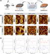
(a) Schematic representation of the fabrication of non-porous thin films by spin coating, followed by a hydrolysis step of the PLA grafts to obtain porous thin films. Topographical AFM images of the (b) non-porous films and (c) porous films after PLA hydrolysis. The white arrows indicate the places where some holes are located. Red (1) and blue (2) lines show the cut-positions to determine the pore dimensions in (d).
Figure 3
Figure 3
TEM images, including the EDX analysis showing sulfur atoms in red and carbon atoms in blue, of the (a) non-porous films, and (b) porous films after PLA hydrolysis. The white arrows indicate the formation of pores.
Figure 4
Figure 4
Normalized optical absorption spectra of (a) non-porous (solid curves) and (b) porous (dashed curves) thin films. Normalized fluorescence spectra (λexc = 480 nm) of (c) non-porous (solid curves) and (d) porous (dashed curves) thin films.
Figure 5
Figure 5
(a) Schematic representation of the PEC used for measuring the photocurrent properties and electrical capacitance. (b) Equivalent circuit model of the ITO–P3HT–PBS heterojunction used to fit EIS data. (c) Photocurrent curves of non-porous P3HT films with different thicknesses and porous films made of P3HT7-g-PLA93 (blue curve), P3HT28-g-PLA72 (green curve), and P3HT60-g-PLA40 (red curve) when irradiated with a LED at 110 mW cm-2 and 530 nm. (d) Photocurrent values after 1 s of LED irradiation of the non-porous and porous films. (e) Capacitances of non-porous and porous films as obtained from EIS, n = 3.
Figure 6
Figure 6
(a) In vitro proliferation assay of HUVECs plated on P3HT, non-porous (solid bars), and porous (frame filler bars) P3HT28-g-PLA72 films up to 120 h. (b) In vitro ROS production test of non-porous (solid bars) and porous (frame filler bars) P3HT28-g-PLA72 films in contact with HUVECs cells in the darkness and after irradiation with a LED light (λexc = 520 nm; 110 mW cm–2) for 2.5 s. Results are shown as mean ± s.e.m. (3 biological replicates, n = 9 samples per each condition, m = 900 cells per each condition) with statistical tests performed by ANOVA with Bonferroni correction at a significance level of ***p < 0.001. (c) Schematic representation of the SECM setup for the quantitative measurement of photo-induced ROS production at the polymer/electrolyte P3HT/PBS interface. (d) Oxidation currents of H2O2 measured at a black platinum working microelectrode at 0.4 V vs Ag/AgCl (KCl 3 M) for P3HT thin films (brown curves), non-porous (green curves), and porous (blue curves) P3HT28-g-PLA72 thin films, n = 3.

References

    1. Sies H.; Jones D. P. Reactive oxygen species (ROS) as pleiotropic physiological signalling agents. Nat. Rev. Mol. Cell Biol. 2020, 21, 363–383. 10.1038/s41580-020-0230-3. - DOI - PubMed
    1. D’Autréaux B.; Toledano M. B. ROS as signalling molecules: mechanisms that generate specificity in ROS homeostasis. Nat. Rev. Mol. Cell Biol. 2007, 8, 813–824. 10.1038/nrm2256. - DOI - PubMed
    1. Shields H. J.; Traa A.; Van Raamsdonk J. M. Beneficial and Detrimental Effects of Reactive Oxygen Species on Lifespan. A Comprehensive Review of Comparative and Experimental Studies. Front. Cell Dev. Biol. 2021, 9, 628157.10.3389/fcell.2021.628157. - DOI - PMC - PubMed
    1. Zimmerman M. C.; Case A. J. Redox biology in physiology and disease. Redox Biol. 2019, 27, 101267.10.1016/j.redox.2019.101267. - DOI - PMC - PubMed
    1. Adhikari A.; Mondal S.; Chatterjee T.; Das M.; Biswas P.; Ghosh R.; Darbar S.; Alessa H.; Althakafy J. T.; Sayqal A.; et al. Redox nanomedicine ameliorates chronic kidney disease (CKD) by mitochondrial reconditioning in mice. Commun. Biol. 2021, 4, 1013.10.1038/s42003-021-02546-8. - DOI - PMC - PubMed