. 2023 Jul 18;4(7):101113.
doi: 10.1016/j.xcrm.2023.101113.

A surgically optimized intraoperative poly(I:C)-releasing hydrogel prevents cancer recurrence

Affiliations

A surgically optimized intraoperative poly(I:C)-releasing hydrogel prevents cancer recurrence

Francois Xavier Rwandamuriye et al. Cell Rep Med. .

Abstract

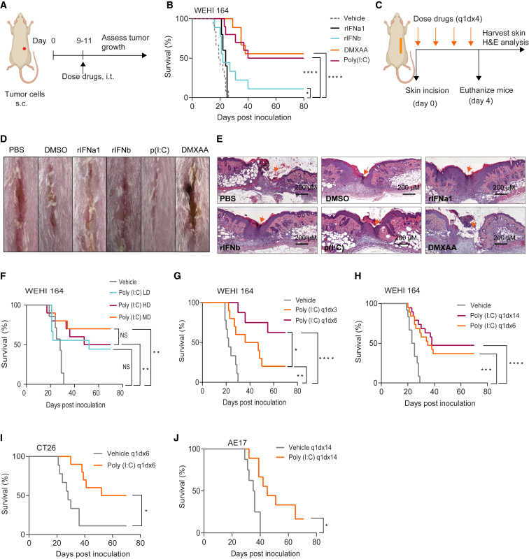
Recurrences frequently occur following surgical removal of primary tumors. In many cancers, adjuvant therapies have limited efficacy. Surgery provides access to the tumor microenvironment, creating an opportunity for local therapy, in particular immunotherapy, which can induce local and systemic anti-cancer effects. Here, we develop a surgically optimized biodegradable hyaluronic acid-based hydrogel for sustained intraoperative delivery of Toll-like receptor 3 agonist poly(I:C) and demonstrate that it significantly reduces tumor recurrence after surgery in multiple mouse models. Mechanistically, poly(I:C) induces a transient interferon alpha (IFNα) response, reshaping the tumor/wound microenvironment by attracting inflammatory monocytes and depleting regulatory T cells. We demonstrate that a pre-existing IFN signature predicts response to the poly(I:C) hydrogel, which sensitizes tumors to immune checkpoint therapy. The safety, immunogenicity, and surgical feasibility are confirmed in a veterinary trial in canine soft tissue tumors. The surgically optimized poly(I:C)-loaded hydrogel provides a safe and effective approach to prevent cancer recurrence.

Keywords: PD-1; Toll-like receptor; cancer; drug delivery; hydrogel; immunotherapy; poly(I:C); surgical oncology; tumor; wound healing.


Conflict of interest statement

Declaration of interests We declare a patent application pertaining to aspects of this work. B.F.K. discloses a financial interest in OncoRes Medical, a company developing QME for use in surgery. W.J.L. declares consultancy for Douglas Pharmaceuticals and MSD and research funding from Douglas Pharmaceuticals, AstraZeneca, and ENA Therapeutics.

Figures

None
Graphical abstract
Figure 1
Figure 1
Design and characterization of a surgically optimized hyaluronic acid-based hydrogel (A) Representative photographs of different hydrogel formulations obtained using a constant percentage of HA polymer (2.5% w/v) and varying amounts of crosslinker (1.8–10 mol %). (B and C) Young’s modulus for different hydrogel formulations with varying amounts of DTPH crosslinker (B) or HA polymer (C). Data are mean ± SEM; n = 3 replicates for each hydrogel. Experiments were performed more than twice in (A) and once in (B) and (C). See also Figure S1A. (D) Scanning electron microscopy (SEM) image of the optimized hydrogel (2.5% w/v HA polymer and 3.5 mol % crosslinker). Scale bar, 5 μM. (E) Quantitative micro-elastography (QME) scan of the optimized hydrogel. Scale bar, 1 mm. Volumetric QME scans were acquired over a 3 × 3 × 2.5 mm (x, y, z) field of view in the center of HA gel disc with a voxel size of 3 × 3 × 2.5 μm (x, y, z). This voxel size resulted in 1 million elasticity measurements in each two-dimensional (2D) image. Experiments were performed once in (E). See also Figure S1B. (F) Representative in vivo fluorescence images for gel degradation. HA hydrogels were labeled with Cy7; n= 3 mice per group. (G) In vivo gel degradation profile. Cy7 signal as total signal (×106 phot/cm2/s). Data are mean ± SD; n = 3 mice per group. Experiments were performed twice in experiments (F) and (G). (H) In vivo mobility of hydrogels. In vivo fluorescence images of free Cy7 resuspended in saline, a non-crosslinked Cy7-labeled HA hydrogel (2.5% w/v HA, non-crosslinked) or the crosslinked hydrogel (2.5% w/v HA, 3.5 mol % DTPH) labeled with Cy7, imaged at 3 h after intraperitoneal injection in mice; n = 2 mice per group. The experiment was performed once.
Figure 2
Figure 2
Prolonged intratumoral poly(I:C) treatment is effective and safe at low dose (A) Experimental design. Mice with established tumors were treated intratumorally (i.t.) with immune adjuvants, daily, for 3, 6, or 14 days, depending on the treatment schedule. (B) Survival curves of WEHI 164-bearing mice treated with poly(I:C) 10 μg/day, DMXAA 50 μg/day, rIFNα 2,000 IU/day, rIFNβ 2,000 IU/day, or vehicle daily for 6 days; n = 8–10 mice per group. (C) Experimental setup for the skin incision model. Mice were dosed with poly(I:C), DMXAA, rIFNα, or rIFNβ in the wound area, daily, for 4 days, using the same doses as in (B); n = 3–5 mice per group. (D and E) Representative photographs of mice showing macroscopic wound healing (D) and H&E staining of skin cross-sections around the wounded area (E) collected on day 4 post-surgery. Scale bar, 200 μm. Magnification, 20×. (F) Survival curves of WEHI 164-bearing mice treated with different doses of poly(I:C), daily, for 3 days, peritumorally, after 50% debulk of the primary tumor; n = 8–10 mice per group. Poly(I:C) doses: 1 μg/day (low dose [LD]), 10 μg/day (medium dose [MD]), or 50 μg/day (high dose [HD]). (G and H) Survival curves of WEHI 164-bearing mice treated with poly(I:C) (10 μg/day), comparing 3 versus 6 days (G) or 6 versus 14 days (H); n = 8–10 mice per group. (I and J) Survival curves of CT26-bearing mice (I) or AE17-bearing mice (J) treated with poly(I:C), i.t., daily, for 6 days; n = 8–10 mice per group. NS, not significant; Poly(I:C), polyinosinic:polycytidylic acid; DMXAA, 5,6-dimethylxantenone-4-acetic acid; rIFNβ, recombinant interferon beta; rIFNα1, recombinant interferon alpha. In (B) and (F)–(J), experiments were performed twice, and statistical analyses were performed using the log rank (Mantel-Cox) test to compare survival. Significance is represented using asterisks as follows: ∗p ≤ 0.05, ∗∗p ≤ 0.005, ∗∗∗p ≤ 0.0005, and ∗∗∗∗p ≤ 0.0001.
Figure 3
Figure 3
An intraoperative poly(I:C)-releasing hydrogel prevents tumor recurrence (A) In vitro cumulative release profile of poly(I:C) from the optimized hydrogel with different concentration of hyaluronidase. Data are mean ± SD; n = 3 technical replicates. The experiment was performed once. (B) Representative in vivo fluorescence images. Top row: Cy7-labeled gel, without poly(I:C), as a control arm. Middle row: Cy7-labeled gel plus unlabeled poly(I:C) as a control arm. Bottom row: Cy7-labeled poly(I:C) encapsulated in the hydrogel; n = 3 mice per group. The experiment was performed twice. (C) In vivo poly(I:C)-Cy7 release profile. Cy7 signal as total signal (×106 phot/cm2/s). Data are mean ± SD; n = 3 mice per group. Statistical comparison was made using one-way ANOVA followed by Tukey’s multiple-comparison test. NS, not significant. (D–F) Efficacy of hydrogel-poly(I:C) implanted in the tumor resection cavity after incomplete tumor resection. (D) Experimental design. A 75% debulk (WEHI 164 tumors) or 90% debulk (CT26 tumors) was performed and 100 μL of hydrogel-loaded poly(I:C) (250 μg) or empty hydrogel was placed in the resection cavity; n = 8–10 mice per group. The experiments were performed twice. (E and F) Survival curves of mice treated with hydrogel-poly(I:C) in WEHI 164 (E) or CT26 (F). In (E) and (F), statistical analyses were performed using the log rank (Mantel-Cox) test to compare survival. Significance is represented using asterisks as follows: ∗p ≤ 0.05, ∗∗p ≤ 0.005, ∗∗∗p ≤ 0.0005, ∗∗∗∗p ≤ 0.0001.
Figure 4
Figure 4
Prolonged poly(I:C) administration induces a transient IFNα response attracting inflammatory myeloid cells and depleting Tregs in the TME (A) Experimental setup for poly(I:C) uptake studies. Mice bearing WEHI 164 tumors were treated with poly(I:C), i.t., daily, for 4 days and a single injection of fluorescein-labeled poly(I:C) (50 μg), i.t., one hour before harvesting tumors for flow cytometry; n = 3 mice per group. (B) Representative t-distributed stochastic neighbor embedding (t-SNE) plots. (C–H) Time-dependent analysis of gene expression in poly(I:C)- or vehicle-treated tumors. (C) Experimental design, and treatment strategy. (D) Heatmap of differentially expressed genes between poly(I:C) and vehicle-treated groups across the different time points; n = 3 mice per group. (E–H) TC-seq analysis was used to cluster genes with similar expression over time. (E and F) Gene expression over time for cluster 1 and cluster 2. (G and H) Top 5 upregulated biological pathways in clusters 1 and 2. In (A)–(H), the experiments were performed once. (I) Experimental design for cytokine blocking studies; n = 8–10 mice per group. The experiments were performed twice. (J) Survival curves of WEHI 164-bearing mice treated with poly(I:C) with or without blocking IFNα or IFNβ, or their receptor IFNAR, or blocking IFNγ. Statistical analyses were performed using the log rank (Mantel-Cox) test to compare survival. Significance is represented using asterisks as follows: ∗p ≤ 0.05, ∗∗p ≤ 0.005, ∗∗∗p ≤ 0.0005, ∗∗∗∗p ≤ 0.0001. (K) Deconvolution analysis of RNA-seq data from Figures 1C and 1D; n = 3 mice per group. (L–R) Mice bearing WEHI 164 tumors were treated with 2 doses of anti-IFNAR1 starting one day prior to 4 daily i.t. poly(I:C) injections. Tumors were harvested at day 4 for flow cytometry analysis; n = 5–6 mice per group. The experiments were performed twice. (L) UMAP showing clustering of cell populations across different treatment groups. (M and N) Representative FACS plots and proportion of inflammatory monocytes (CD11b+Ly-6ChighLy-6G). (O and P) Representative FACS plots and proportion of Tregs (CD4+FoxP3+). (Q and R) Representative FACS plots and proportion of PD-L1+ macrophages (CD11bhighF4/80+PD-L1+). Data are mean ± SD; n = 5–6 biologically independent samples per group. In (L)–(R), statistical analyses were performed using one-way ANOVA followed by Tukey’s multiple-comparison test. NS, not significant. (S) Survival curves of WEHI 164-bearing mice treated with poly(I:C) with or without anti-CD4 or anti-CD8α cell depleting monoclonal antibodies; n = 8–10 mice per group. The experiments were performed twice. Statistical analyses were performed using the log rank (Mantel-Cox) test to compare survival. Significance is represented using asterisks as follows: ∗p ≤ 0.05, ∗∗p ≤ 0.005, ∗∗∗p ≤ 0.0005, ∗∗∗∗p ≤ 0.0001. See also Figures S3, S4, and S6A.
Figure 5
Figure 5
A pre-existing IFN gene signature predicts response to poly(I:C) hydrogel which sensitizes tumors to PD-1 and CTLA-4 blockade (A and B) Efficacy of poly (I:C) in tumors derived from TLR3 KO WEHI 164 cells. (A) Experimental design and treatment strategy. (B) Survival curves of wild-type WEHI 164- or WEHI 164 TLR3 KO-bearing mice treated with poly(I:C); n = 8–10 mice per group. The experiments were performed twice. See also Figures S5A and S5B. (C and D) RNA-seq for responders versus non-responders. (C) Experimental design, and treatment strategy. Partially resected tumors were kept in RNAlater for subsequent RNA extraction and sequencing. Treated mice were assigned to responder and non-responder groups depending on tumor outgrowth; n = 4–6 mice per group. The experiment was performed once in (D). (D) GSEA top hallmark gene sets in responsive versus nonresponsive tumors. IL2, interleukin-2; IL6, interleukin-6; JAK, Janus kinase; STAT3/5, signal transducer and activator of transcription 3/5. See also Figures S5C–S5F. (E–G) Combination of hydrogel poly(I:C) with anti-PD-1 or anti-CTLA-4. (E) Experimental design and treatment strategy. (F and G) Survival of CT26-bearing mice treated with poly(I:C) hydrogel in combination with anti-PD-1 (F) or anti-CTLA-4 (G); n = 8–10 mice per group. The experiments were performed twice. In (B), (F), and (G), statistical analyses were performed using the log rank (Mantel-Cox) test to compare survival. Significance is represented using asterisks as follows: ∗p ≤ 0.05, ∗∗p ≤ 0.005, ∗∗∗p ≤ 0.0005, ∗∗∗∗p ≤ 0.0001.
Figure 6
Figure 6
Safety, feasibility, and immunostimulatory potential of the poly(I:C) hydrogel in a canine trial (A) Experimental design. Canine patients received intraoperative KLH/poly(I:C) hydrogel, containing 0.2 mg poly(I:C) and 1 mg KLH, at the time of surgery. Patient blood samples were taken directly prior to surgery, and at 2 weeks post-surgery. See STAR Methods for canine patient characteristics. (B) Representative photographs of hydrogel application during surgical resection of a soft tissue tumor in a canine patient. (C and D) Representative FACS plots for KLH-specific proliferation (C) or Ki67 and granzyme B expression (D) in CD4+ and CD8+ T cells from canine patient peripheral blood mononuclear cells (PBMCs) collected 2 weeks post-surgery. PBMCs were restimulated in complete media with or without KLH (20 μg/mL) for 72 h prior to flow cytometry analysis. CFSE, carboxyfluorescein succinimidyl ester. See also Figure S6B. (E) Quantification of pro-inflammatory cytokines in culture supernatants taken 48 h after ex vivo restimulation of PBMCs. Representative data are from one canine patient. Data are from one biological independent subject. In (E), two technical replicates were performed.

Similar articles

Cited by

References

    1. Wyld L., Audisio R.A., Poston G.J. The evolution of cancer surgery and future perspectives. Nat. Rev. Clin. Oncol. 2015;12:115–124. doi: 10.1038/nrclinonc.2014.191. - DOI - PubMed
    1. Mahvi D.A., Liu R., Grinstaff M.W., Colson Y.L., Raut C.P. Local cancer recurrence: The realities, challenges, and opportunities for new therapies. CA A Cancer J. Clin. 2018;68:488–505. doi: 10.3322/caac.21498. - DOI - PMC - PubMed
    1. Eggermont A.M.M., Blank C.U., Mandala M., Long G.V., Atkinson V., Dalle S., Haydon A., Lichinitser M., Khattak A., Carlino M.S., et al. Adjuvant pembrolizumab versus placebo in resected stage iii melanoma. N. Engl. J. Med. 2018;378:1789–1801. doi: 10.1056/NEJMoa1802357. - DOI - PubMed
    1. Forde P.M., Chaft J.E., Smith K.N., Anagnostou V., Cottrell T.R., Hellmann M.D., Zahurak M., Yang S.C., Jones D.R., Broderick S., et al. Neoadjuvant pd-1 blockade in resectable lung cancer. N. Engl. J. Med. 2018;378:1976–1986. doi: 10.1056/NEJMoa1716078. - DOI - PMC - PubMed
    1. Cloughesy T.F., Mochizuki A.Y., Orpilla J.R., Hugo W., Lee A.H., Davidson T.B., Wang A.C., Ellingson B.M., Rytlewski J.A., Sanders C.M., et al. Neoadjuvant anti-pd-1 immunotherapy promotes a survival benefit with intratumoral and systemic immune responses in recurrent glioblastoma. Nat. Med. 2019;25:477–486. doi: 10.1038/s41591-018-0337-7. - DOI - PMC - PubMed

Publication types