. 2023 Jul 25;82(4):317-332.
doi: 10.1016/j.jacc.2023.05.031.

Role of Human Epicardial Adipose Tissue-Derived miR-92a-3p in Myocardial Redox State

Affiliations

Role of Human Epicardial Adipose Tissue-Derived miR-92a-3p in Myocardial Redox State

Maria Cristina Carena et al. J Am Coll Cardiol. .

Abstract

Background: Visceral obesity is directly linked to increased cardiovascular risk, including heart failure.

Objectives: This study explored the ability of human epicardial adipose tissue (EAT)-derived microRNAs (miRNAs) to regulate the myocardial redox state and clinical outcomes.

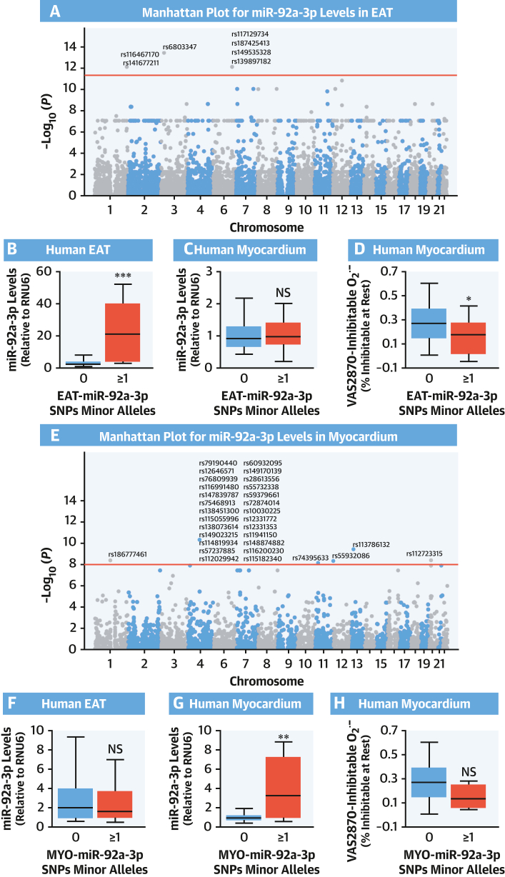
Methods: This study screened for miRNAs expressed and released from human EAT and tested for correlations with the redox state in the adjacent myocardium in paired EAT/atrial biopsy specimens from patients undergoing cardiac surgery. Three miRNAs were then tested for causality in an in vitro model of cardiomyocytes. At a clinical level, causality/directionality were tested using genome-wide association screening, and the underlying mechanisms were explored using human biopsy specimens, as well as overexpression of the candidate miRNAs and their targets in vitro and in vivo using a transgenic mouse model. The final prognostic value of the discovered targets was tested in patients undergoing cardiac surgery, followed up for a median of 8 years.

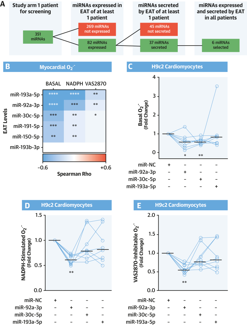
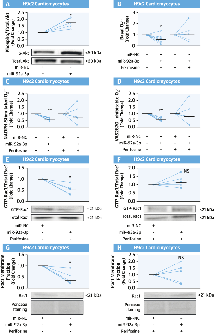
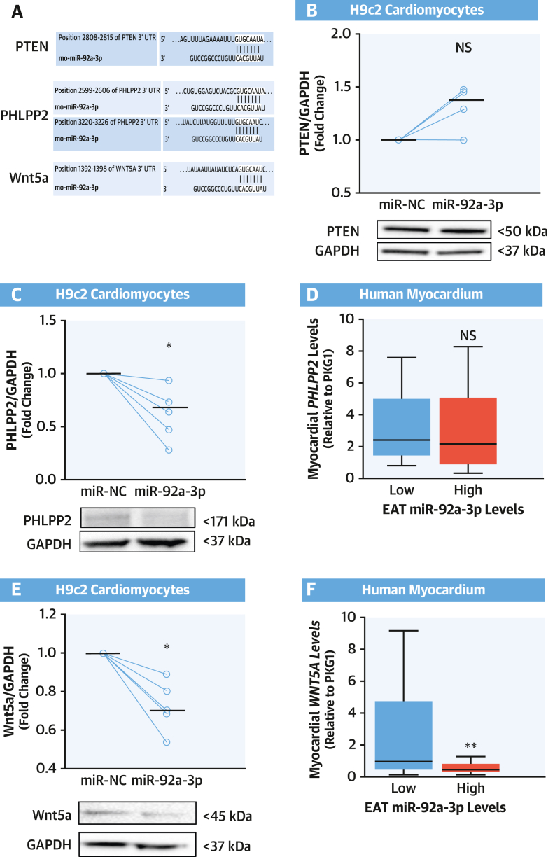
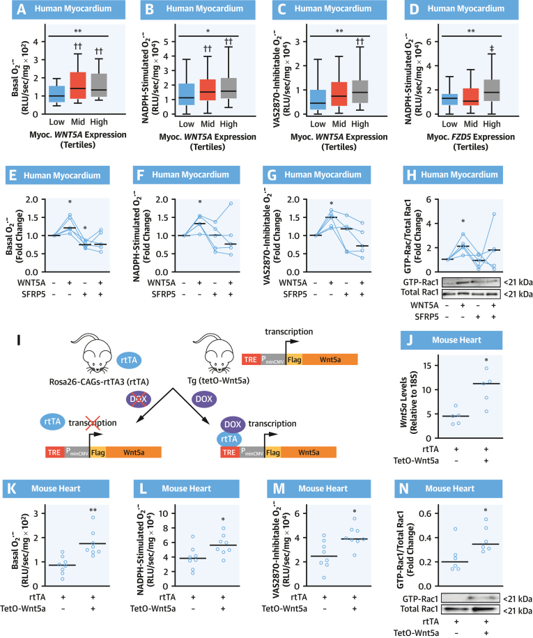
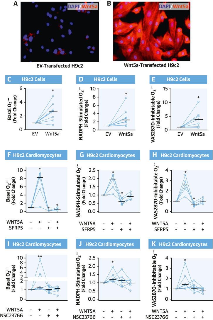
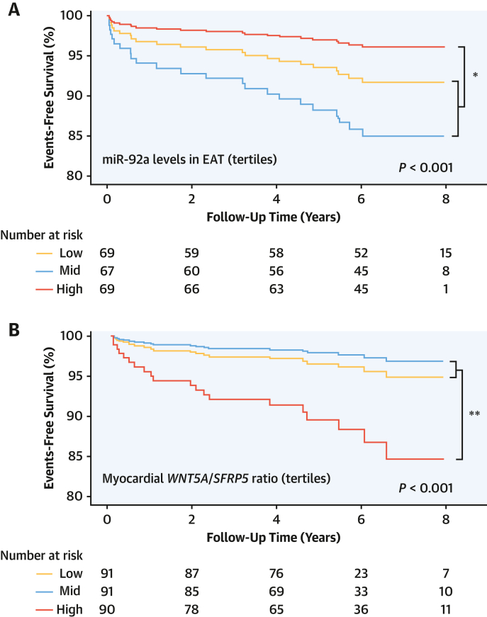
Results: EAT miR-92a-3p was related to lower oxidative stress in human myocardium, a finding confirmed by using genetic regulators of miR-92a-3p in the human heart and EAT. miR-92a-3p reduced nicotinamide adenine dinucleotide phosphate (NADPH)-oxidase-derived superoxide (O2.-) by targeting myocardial expression of WNT5A, which regulated Rac1-dependent activation of NADPH oxidases. Finally, high miR-92a-3p levels in EAT were independently related with lower risk of adverse cardiovascular events.

Conclusions: EAT-derived miRNAs exert paracrine effects on the human heart. Indeed miR-92a-3p suppresses the wingless-type MMTV integration site family, member 5a/Rac1/NADPH oxidase axis and improves the myocardial redox state. EAT-derived miR-92a-3p is related to improved clinical outcomes and is a rational therapeutic target for the prevention and treatment of obesity-related heart disease.

Keywords: Wnt5a signaling; epicardial adipose tissue; microRNAs; myocardial NADPH oxidase activity; myocardial oxidative stress.


Conflict of interest statement

Funding Support and Author Disclosures This study was supported by a Marie Skłodowska-Curie Early Stage Researcher fellowship to Dr Carena; the CATCH ME (Characterizing Atrial fibrillation by Translating its Causes into Health Modifiers in the Elderly) consortium (grant number 633196); the British Heart Foundation (FS/16/15/32047, RG/F/21/110040 and CH/F/21/90009 to Dr Antoniades; CH/16/1/32013 to Dr Channon; CH/12/3/29609 to Dr Casadei); Oxford BHF Centre of Research Excellence RE/18/3/34214, the Oxford NIHR Biomedical Research Centre, the National Institute for Health Research Oxford Biomedical Research Centre, and the Novo Nordisk Foundation (NNF15CC0018486) to Dr Antoniades. Dr Antoniades has had consultancy agreements with Mitsubishi Tanabe and Silence Therapeutics; has received grants from Sanofi and Novo Nordisk; is the Chair of the British Atherosclerosis Society; and has received honoraria from Amarin and Covance. Drs Antoniades and Channon are founders, shareholders, and directors of Caristo Diagnostics. Dr Casadei is the past president of the European Society of Cardiology. All other authors have reported that they have no relationships relevant to the contents of this paper to disclose.

Figures

None
Graphical abstract
Figure 1
Figure 1
EAT-Derived miR-92a-3p and Myocardial O2.– Production (A) Flowchart showing the screening process to identify microRNAs (miRNAs) expressed and released by epicardial adipose tissue (EAT). (B) Heatmap showing the Spearman correlation coefficient between the EAT levels of the 6 identified miRNAs and the basal, nicotinamide adenine dinucleotide phosphate (NADPH)-stimulated, and VAS2870-inhibitable superoxide (O2.–) production in the human myocardium (n = 56). ∗P < 0.05, ∗∗P < 0.01, ∗∗∗P < 0.001, ∗∗∗∗P < 0.0001. (C to E) Basal, NADPH-stimulated, and VAS2870-inhibtable O2.– generation in differentiated H9c2 cardiomyocytes transfected with an miRNA mimic negative control (miR-NC) or miR-30c-5p, miR-92a-3p, and miR-193a-5p mimic (n = 8). Lines on scatterplots represent medians. ∗P < 0.05, ∗∗P < 0.01 vs control by Wilcoxon signed-rank test.
Figure 2
Figure 2
SNPs Affecting EAT miR-92a-3p and Myocardial Redox State Manhattan plots representing single-nucleotide polymorphisms (SNPs) associated with miR-92a-3p levels in EAT (A) and myocardium (E). Seven SNPs were significantly associated with high miR-92a-3p levels in EAT (EAT-miR-92a-3p) and 31 with high levels of miR-92a-3p in the myocardium (MYO-miR-92a-3p). The presence of any SNP from EAT-miR-92a-3p was associated with higher miR-92a-3p levels only in EAT, not in the myocardium (B, n = 149; C, n = 265), whereas the presence of any MYO-miR-92a-3p SNP was associated with higher miR-92a-3p levels only in the myocardium, not in EAT (G, n = 265; F, n = 149). The presence of any EAT-miR-92a-3p SNP led to a statistically significant reduction of myocardial superoxide production (D and H, n = 196). In B to D and F to H, data are presented as median (25th-75th percentile). ∗P < 0.05, ∗∗P < 0.01, ∗∗∗P < 0.001 by the Mann-Whitney U test. NS = not significant; other abbreviations as in Figure 1.
Figure 3
Figure 3
miR-92a-3p Modulates O2.– Production in Cardiomyocytes Through Akt (A) Transfection of differentiated H9c2 cardiomyocytes with miR-92a-3p mimic increased the phosphorylation of Akt protein kinase at Ser473 compared with an miR-NC (n = 5). (B to D) miR-92a-3p expression in differentiated H9c2 cardiomyocytes suppressed NADPH-stimulated and VAS2870-inhibtable O2.– compared with miR-NC, an effect abolished in the presence of the Akt inhibitor perifosine (n = 5-8). (E to H) Fold change of activated Rac1 (measured as ratio of guanosine triphosphate [GTP]-Rac1:total Rac1) and of Rac1 membrane translocation in H9c2 cardiomyocytes transfected with either miR-NC or a miR-92a-3p mimic ± Akt inhibitor perifosine (n = 5). Lines on scatterplots represent medians. ∗P < 0.05, ∗∗P < 0.01 vs controls by Wilcoxon signed-rank test. Abbreviations as in Figure 1.
Figure 4
Figure 4
miR-92a-3p Downregulates Protein Levels of Wnt5a in Cardiomyocytes (A) Prediction of miR-92a-3p binding to phosphatase and tensin homolog (PTEN), PH domain and leucine-rich repeat protein phosphatase 2 (PHLPP2), and wingless-type MMTV integration site family, member 5a (Wnt5a) 3′-untranslated regions (UTRs) assessed by using TargetScan software version 7.2 (TargetScan). Transfection of H9c2 cardiomyocytes with miR-92a-3p mimic resulted in the reduction of protein levels of PHLPP2 (C) and Wnt5a (E), but not PTEN (B), compared with the miR-NC (n = 4-6). (D and F) Patients with high levels (above the median) of miR-92a-3p in EAT had lower myocardial expression of WNT5A (but not PPLPP2) (n = 71). Data are presented as median (25th-75th percentile). Lines on scatterplots represent medians. ∗P < 0.05 vs control by Wilcoxon signed-rank test. ∗∗P < 0.01 by Mann-Whitney U test. GAPDH = glyceraldehyde 3-phosphate dehydrogenase; other abbreviations as in Figures 1 and 3.
Figure 5
Figure 5
WNT5A Increases O2.– Generation in Human and Murine Myocardium (A to D) Basal, NADPH-stimulated, and VAS2870-inhibtable O2.– production in human myocardium according to tertiles of WNT5A or FZD5 expression in myocardium (n = 189 in A to C; n = 181 in D). Data are presented as median (25th-75th percentile). ∗P < 0.05, ∗∗P < 0.01 by Kruskal-Wallis test; ††P < 0.01 vs low tertile by Dunn’s test corrected for multiple tests. (E to H) O2.– production and fold change of activated Rac1 in human myocardium in presence/absence of WNT5A and SFRP5 (n = 5). ∗P < 0.05 vs control by Wilcoxon signed paired rank test. (I) Breeding scheme for inducible expression of FLAG-tagged Wnt5a. (J) Doxycycline (DOX) treatment induces marked Wnt5a overexpression in Wnt5a+/rtTA+ hearts (n = 5). O2.– production (K to M) and Rac1 activation (N) in hearts of DOX-treated mice (n = 6-8 per group). Lines on scatterplots represent median values. ∗P <0.05, ∗∗P < 0.01 vs control by unpaired t test. Myoc = myocardial; PminCMV = promoter; RLU = relative light unit; rtTA = reverse tetracycline-controlled transactivator; TRE = tetracycline-response element; other abbreviations as in Figures 1 and 4.
Figure 6
Figure 6
Wnt5a Increases O2.– Generation in Cardiomyocytes Through Rac1-Mediated NOX Activity (A and B) Overexpression of FLAG-tagged Wnt5a in H9c2 cells was evaluated by immunofluorescence with anti-FLAG antibody (red) and compared with H9c2 cells transfected with an empty vector (EV); nuclei were stained with 4′,6-diamidino-2-phenylindole (DAPI) (blue). (C to E) Basal, NADPH-stimulated, and VAS2870-inhibtable O2.– in H9c2 cells overexpressing or not FLAG-tagged Wnt5a (n = 8). (F to H) O2.– production in H9c2 cardiomyocytes in the presence/absence of WNT5A and SFRP5 (n = 5). (I to K) O2.– production in H9c2 cardiomyocytes treated with or without WNT5A and NSC23766, a specific RAC1 inhibitor (n = 5-7). Lines represent medians. ∗P < 0.05, ∗∗P < 0.01 vs control by Wilcoxon signed-rank test. Abbreviations as in Figures 1 and 4.
Figure 7
Figure 7
Association of EAT miR-92a-3p Levels With Risk of Cardiovascular Events Prognostic value of epicardial adipose tissue (EAT) miR-92a-3p levels (A) and myocardial WNT5A/SFRP5 levels (B) for the composite outcome of cardiac mortality, nonfatal myocardial infarction, and nonfatal stroke. The P values are calculated from Cox regression after adjusting for age, sex, hypertension, body mass index, and diabetes. ∗P < 0.05, ∗∗P < 0.01 for highest tertile vs pooled mid and lowest tertiles. Other abbreviations as in Figures 1 and 4.
Central Illustration
EAT miR-92a-3p, Myocardial Redox State, and Clinical Outcomes Epicardial adipose tissue (EAT) expresses and releases miR-92a-3p that decreases superoxide production in cardiomyocytes possibly by targeting wingless-type MMTV integration site family, member 5a (Wnt5a). Wnt5a induces guanosine triphosphate (GTP) activation of Rac1 that translocates to the cell membrane together with other cytosolic regulatory subunits to form an active enzymatic complex containing nicotinamide adenine dinucleotide phosphate (NADPH) oxidases (eg, NOX2), which catalyzes the production of superoxide (O2.–). High EAT miR-92a-3p is associated with lower cardiovascular risk.

Comment in

References

    1. Roth G.A., Mensah G.A., Johnson C.O., et al. Global burden of cardiovascular diseases and risk factors, 1990-2019: update from the GBD 2019 study. J Am Coll Cardiol. 2020;76:2982–3021. - PMC - PubMed
    1. Akoumianakis I., Antoniades C. The interplay between adipose tissue and the cardiovascular system: is fat always bad? Cardiovasc Res. 2017;113:999–1008. - PubMed
    1. Oikonomou E.K., Antoniades C. The role of adipose tissue in cardiovascular health and disease. Nat Rev Cardiol. 2019;16:83–99. - PubMed
    1. Antonopoulos A.S., Margaritis M., Verheule S., et al. Mutual regulation of epicardial adipose tissue and myocardial redox state by PPAR-γ/adiponectin signalling. Circ Res. 2016;118:842–855. - PMC - PubMed
    1. Winkle M., El-Daly S.M., Fabbri M., Calin G.A. Noncoding RNA therapeutics—challenges and potential solutions. Nat Rev Drug Discov. 2021;20:629–651. - PMC - PubMed

Publication types